
Introduction:
If you have been told that you have a cataract, you have probably heard several unfamiliar terms during your eye consultation. One term that comes up again and again is phacoemulsification, often simply called phaco. It sounds complicated, but the basic idea is surprisingly straightforward: the cloudy natural lens inside your eye is gently broken into tiny pieces using ultrasound energy, removed through a small opening, and replaced with a clear artificial lens called an intraocular lens (IOL). Today, small-incision phacoemulsification with foldable IOL implantation is one of the predominant approaches to cataract surgery, particularly where the necessary surgical technology and expertise are available.
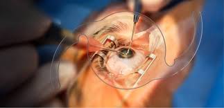
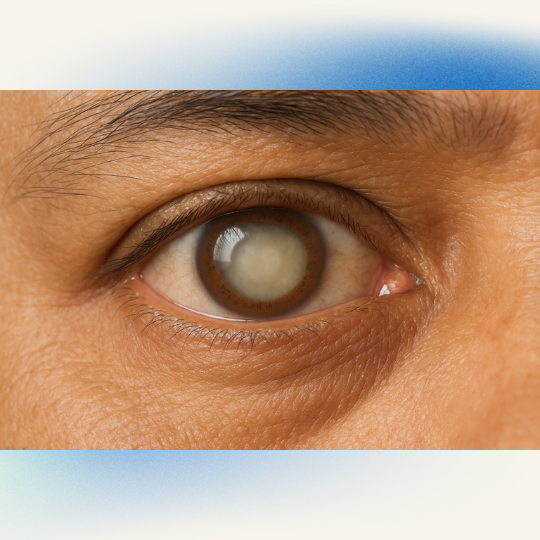
Why does this matter? A cataract can gradually turn what was once a clear view into something like looking through a fogged window. Colours may appear dull, lights can become uncomfortable, reading may become difficult, and driving—especially at night—can become increasingly challenging. Cataracts do not disappear through eye exercises, dietary supplements, or stronger glasses; when the cataract significantly affects vision and daily activities, surgery is the definitive treatment.
The good news is that modern cataract surgery has changed dramatically. Earlier techniques often required larger incisions, whereas modern phacoemulsification generally uses a small incision and a foldable IOL. The smaller approach can reduce surgically induced astigmatism and support faster visual rehabilitation compared with larger-incision techniques.
What Is Phacoemulsification?
Phacoemulsification removes cataracts by using ultrasound energy to break up the cloudy natural lens into small fragments, which the surgeon then aspirates and removes. The surgeon places an artificial intraocular lens (IOL) inside the eye to restore its focusing function. The procedure usually requires only a small corneal incision, and the surgeon preserves the natural lens capsule whenever possible to support the implanted IOL.
Think of the cloudy lens as a small, hardened piece of material sitting inside a delicate container. Instead of trying to pull that entire piece through a large opening, phacoemulsification allows the surgeon to break the lens into manageable fragments and remove them through a much smaller incision. This is one of the major reasons the technique became such an important development in modern cataract surgery. The technology traces its modern origins to Charles Kelman’s introduction of phacoemulsification in 1967, after which advances in IOL design, surgical instruments and ophthalmic viscosurgical devices helped make small-incision cataract surgery increasingly practical.
Phacoemulsification should not be confused with laser eye surgery such as LASIK. Phaco uses ultrasound energy inside the eye to remove the cataractous lens, while LASIK reshapes the cornea to correct refractive errors such as myopia, hyperopia and astigmatism. NHS patient information specifically describes phacoemulsification as an ultrasound-based process and notes that it does not involve lasers.
How Phacoemulsification Works:
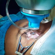
During the procedure, the surgeon first creates a carefully planned small opening in the cornea. The surgeon opens the front portion of the lens capsule in a step called capsulorrhexis and uses fluid to separate and mobilise the lens nucleus. The surgeon then uses a phacoemulsification handpiece with an ultrasonically driven tip to break the cataract into small fragments. The surgical system simultaneously aspirates the fragmented material from the eye.
Once the cloudy lens material has been removed, the surgeon places a suitable artificial lens, usually a foldable IOL, into the remaining capsular bag. The goal is not simply to remove the cataract but to create a stable optical system that provides the best practical visual outcome for the individual patient. Lens selection can therefore be an important part of the overall cataract-surgery discussion.
Why Is Phacoemulsification Used for Cataract Surgery?
The main reason for performing phacoemulsification is to remove a cataract that is interfering with useful vision. A cataract occurs when the normally transparent natural lens becomes cloudy. As the opacity progresses, light does not pass cleanly through the lens, producing symptoms such as blurred vision, glare, reduced contrast and difficulty seeing in low-light conditions. Cataract surgery becomes appropriate when the visual impairment begins to interfere with the patient’s needs and activities rather than simply because a cataract happens to be visible during an examination.
This distinction is important. Not every person with an early cataract needs immediate surgery. Someone may have a mild lens opacity but still read comfortably, work normally and drive safely. Another person with a similar-looking cataract may struggle significantly with night driving, reading or recognising faces. The decision therefore depends on the individual’s visual function, symptoms, eye health, lifestyle and the ophthalmologist’s assessment.
The American Academy of Ophthalmology identifies decline in visual function that no longer meets a person’s visual needs as the primary indication for cataract surgery. Doctors may recommend cataract removal when the cloudy lens interferes with diagnosing or treating diseases at the back of the eye. They may also recommend surgery when the lens causes complications such as inflammation or glaucoma.
When Cataract Surgery May Be Recommended
You may want to discuss cataract surgery with an ophthalmologist if you notice persistent difficulty with activities that previously felt routine. Common complaints include blurred or hazy vision, increased glare from headlights, difficulty reading small print, faded colours and needing brighter light for close work. Some people also notice that their spectacle prescription seems to change repeatedly without giving them the clear vision they expect.
The important question is not simply, “How mature is my cataract?” A better question is, “How much is my cataract affecting my quality of life and visual function?” Your ophthalmologist can evaluate the cataract along with the cornea, retina, optic nerve, eye pressure and other factors before recommending surgery.
How Does Phacoemulsification Differ From Traditional Cataract Surgery?
Traditional cataract extraction techniques have played an important role in restoring sight, and different methods remain useful in different circumstances. In extracapsular cataract extraction (ECCE), the surgeon removes the lens nucleus through a comparatively larger incision. Manual small-incision cataract surgery, or MSICS, also remains widely used, particularly in settings where it offers practical and economic advantages.
Phacoemulsification takes a different approach. Instead of extracting the lens nucleus as one large piece, the surgeon uses ultrasound energy to divide and emulsify it, allowing aspiration through a smaller incision. Modern phacoemulsification procedures commonly use a foldable IOL and allow patients to undergo surgery on an outpatient basis. The AAO notes that phacoemulsification has advantages associated with small incisions, including less surgically induced astigmatism and the ability to incorporate different approaches to astigmatism management and speciality IOLs.
Phacoemulsification vs. Manual Small-Incision Cataract Surgery
| Feature | Phacoemulsification | Manual Small-Incision Cataract Surgery |
|---|---|---|
| Cataract removal | Ultrasound fragmentation and aspiration | Manual removal through a larger tunnel |
| Incision | Generally very small | Generally larger |
| Ultrasound energy | Used | Not required |
| IOL options | Foldable IOLs commonly used | IOL selection depends on the surgical approach. |
| Technology requirement | Higher | Lower |
| Cost considerations | Can be higher because of equipment and consumables | Often more economical |
| Use in difficult cases. | Frequently used, but the technique depends on the cataract and eye. | May be preferred in selected situations |
There is no universal rule that one technique is perfect for every patient. Cataract density, corneal health, pupil size, zonular stability, previous eye surgery and other conditions can influence the surgical plan. In some resource-limited settings, MSICS remains particularly important because it is less dependent on expensive technology.
What Happens Before Phacoemulsification?
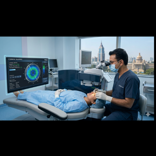
Good cataract surgery starts well before the patient enters the operating room. The preoperative assessment allows the ophthalmologist to determine whether cataract surgery is appropriate and to identify conditions that may affect the expected outcome. The eye is examined carefully, and measurements are taken to help calculate the appropriate IOL power. The NHS also recommends specialist examination and eye measurements before cataract surgery.
Your surgeon may assess visual acuity, the cornea, the anterior chamber, the pupil, the lens, the retina and the optic nerve. Depending on your age, medical history, and eye condition, your doctor may recommend additional investigations. Biometry plays an important role because your doctor selects the appropriate IOL power based on your eye’s dimensions and optical characteristics.
This is also the stage when you should ask questions. Do you want the best possible distance vision? Are you comfortable using reading glasses? Do you have astigmatism? Have you had previous retinal or corneal surgery? Are there other eye conditions that could limit vision even after the cataract is removed? These questions help the surgeon determine which lens and surgical strategy are appropriate.
Step-by-Step Phacoemulsification Procedure
A typical phacoemulsification procedure involves several carefully controlled microsurgical steps. The exact sequence and technique can vary according to the surgeon, the patient’s anatomy and the density of the cataract. Modern cataract surgery commonly includes creation of a small incision, capsulorrhexis, hydrodissection, nuclear fragmentation, removal of remaining lens material and implantation of an IOL.
The patient is usually awake during surgery under local anaesthesia, although the exact anaesthesia plan depends on the individual. Cataract surgery is commonly performed as day surgery under local anaesthesia and usually takes around 30 minutes. However, the actual operating time may vary depending on the patient’s condition and the complexity of the case.
Making the Small Incision
The surgeon creates a small incision in the cornea to allow microsurgical instruments to enter the eye. The surgeon creates the incision to provide access while maintaining the stability of the anterior chamber and minimising changes to the corneal shape. Modern phacoemulsification often requires no sutures because the surgeon can create a self-sealing incision. However, the surgeon may occasionally use sutures or other closure methods when necessary.
The surgeon then creates a controlled opening in the front of the lens capsule. This circular opening provides access to the cataract while helping preserve the capsule that will later support the IOL. The surgeon uses hydrodissection to separate the lens material and facilitate its controlled movement during fragmentation.
Breaking and Removing the Cataract
The phaco handpiece uses ultrasound energy to break the cataract into smaller pieces. Surgical techniques can include approaches such as divide and conquer or various forms of phaco-chop, with the choice depending on the surgeon’s experience, the cataract and the eye’s characteristics. The fragments are then aspirated from the eye through the surgical system.
After removing the nucleus, the surgeon carefully removes the remaining cortical material. The surgeon then prepares the capsular bag for the artificial lens and inserts the foldable IOL, positioning it appropriately inside the eye. Finally, the ophthalmologist ensures that the incision is secure and removes the ophthalmic viscosurgical device material used during surgery.
Choosing the Right Intraocular Lens
Removing the cataract solves one problem, but selecting the right intraocular lens is another important part of the process. An IOL replaces the focusing function of the natural lens after cataract removal. Doctors select different IOL designs based on the patient’s eye measurements, visual goals, corneal astigmatism, and other clinical factors.
A monofocal IOL generally provides a primary focus, often for distance, while glasses may still be needed for reading. Other lens technologies may offer different ranges of vision or address astigmatism, but not every lens is appropriate for every eye. The AAO notes that refractive cataract surgery, including astigmatism management and speciality IOL implantation, may reduce dependence on glasses for some patients.
It is tempting to think that the most expensive lens must automatically be the best lens. That is not necessarily true. The right lens is the one that fits your eye and your visual priorities—not simply the one with the longest list of features. A detailed discussion with your cataract surgeon can help you understand the expected benefits, limitations and potential trade-offs.
Benefits of Phacoemulsification
One of the biggest advantages of phacoemulsification is its small-incision approach. Compared with larger-incision cataract extraction, this can reduce surgically induced astigmatism and support faster visual rehabilitation. The technique also makes it possible to implant foldable IOLs through the small incision.
Another benefit is that the cataract is removed in controlled fragments rather than requiring extraction of the entire lens nucleus through a large incision. Modern phaco machines allow the surgeon to manage ultrasound power, aspiration and fluidics according to the requirements of the case. Some systems also use pulse or burst modes that can help reduce total ultrasound energy during lens removal.
Cataract surgery is commonly performed as day surgery, allowing many patients to return home on the same day. However, same-day discharge does not mean that recovery happens instantly. Your eye still needs time to heal, and your surgeon’s postoperative instructions remain essential.
Is Phacoemulsification Safe?
Phacoemulsification is an established cataract-surgery technique, but no eye operation is completely free of risk. Cataract surgery requires microsurgical precision and the ability to respond appropriately if unexpected problems occur. The AAO emphasises that cataract surgery requires specialist ophthalmic training, judgement and experience.
Potential complications can include infection, inflammation, changes in eye pressure, corneal swelling, posterior capsule rupture, retinal complications and other problems. The individual risk depends on factors such as cataract density, eye anatomy, previous surgery and coexisting eye disease. Some complications are uncommon, but discussing them before surgery is still important because informed consent is part of good medical care.
Modern techniques and careful surgical planning have helped improve cataract surgery outcomes. The AAO reports that randomised clinical trials have found phacoemulsification to have better uncorrected distance visual acuity and lower rates of certain surgical complications compared with some larger-incision cataract extraction approaches.
Recovery After Phacoemulsification
Recovery varies from one patient to another. Many patients notice improved vision soon after surgery, but their vision may initially appear blurred, and the eye may feel mildly uncomfortable or sensitive to light. The eye needs time to settle, and the final visual result can take longer than the first improvement you notice.
Doctors commonly prescribe postoperative eye drops and schedule follow-up appointments to monitor the eye’s healing and recovery. The NHS notes that patients generally return home the same day after cataract surgery and that most people recover quickly, while specific recovery instructions depend on the individual case.
During recovery, it is important not to assume that feeling better means the eye has completely healed. Avoid rubbing or pressing on the operated eye, use prescribed medication exactly as directed and follow your surgeon’s advice regarding bathing, exercise, driving, work and other activities.
Possible Risks and Complications
Although modern cataract surgery has a strong safety record, complications can occur. During surgery, possible problems can include difficulty maintaining the stability of the lens capsule, posterior capsule rupture, retained lens fragments, bleeding or damage to structures within the eye. After surgery, patients can experience inflammation, elevated eye pressure, infection, corneal oedema or retinal complications.
Another condition patients sometimes hear about is posterior capsule opacification (PCO). This is not the cataract growing back. Instead, cells can cause clouding of the capsule behind the IOL after cataract surgery. The surgeon deliberately preserves the capsule during surgery because it provides support for the IOL. Surgical techniques also aim to reduce factors that may contribute to later capsule opacification.
A sudden major decrease in vision, increasing severe pain, marked redness, significant swelling, flashes, a sudden increase in floaters or a curtain-like shadow after surgery should not simply be watched at home. Contact your eye-care team promptly because some postoperative complications require urgent assessment.
Phacoemulsification for Different Types of Cataracts
Not every cataract behaves in exactly the same way. Some are relatively soft and early, while others become very dense and hard. Dense cataracts can require more advanced surgical planning because the surgeon may need to manage the amount of ultrasound energy, maintain good visualisation and protect delicate ocular tissues.
Cataract surgery can also be more complex when the pupil does not dilate well, the zonules supporting the lens are weak, the cornea is cloudy or the eye has undergone previous surgery. The surgical approach may therefore be customised rather than following one identical recipe for every patient. AAO EyeWiki describes how factors such as corneal opacity and cataract density can influence visualisation and the choice of surgical technique.
This is one reason why searching online for a single “best cataract surgery technique” can be misleading. The best approach depends on the eye in front of the surgeon.
Phacoemulsification and Other Eye Conditions
Patients sometimes assume that cataract surgery is a completely isolated procedure, but the rest of the eye matters. Conditions involving the retina, cornea, glaucoma or previous vitreoretinal surgery can influence planning. For example, cataract removal may sometimes be combined with vitreoretinal surgery when both problems require treatment.
In patients with previous vitrectomy, cataract surgery can present additional surgical challenges because the normal fluid dynamics and support within the eye may have changed. EyeWiki notes that phacoemulsification is generally preferred after previous pars plana vitrectomy while also describing the special considerations involved in these cases.
This is why a comprehensive preoperative examination is so valuable. Improving the cloudy lens does not automatically correct every cause of poor vision. If the retina, optic nerve or cornea has another significant problem, the surgeon will explain how that may affect the expected visual outcome.
How Much Does Phacoemulsification Cost?
The cost of phacoemulsification cataract surgery varies considerably depending on the city, hospital, surgeon, diagnostic testing, type of IOL, technology used, anaesthesia and postoperative care. There is no single national price that applies to every patient. Even within the same hospital, the total package can vary depending on the lens selected and the complexity of the procedure.
The IOL can be a particularly important component of the overall cost. A basic monofocal lens and a premium lens designed for additional visual goals may have very different prices. Additional investigations or treatment for other eye conditions can also affect the final bill.
When comparing cataract surgery prices, avoid comparing only the headline number. Ask what the package includes: surgeon fees, facility charges, investigations, IOL cost, medications, follow-up visits and management of unexpected issues. A transparent cost discussion helps you understand what you are actually paying for.
How to Prepare for Cataract Surgery
Preparation begins with a detailed consultation rather than with the operation itself. Tell your ophthalmologist about previous eye surgery, existing eye conditions and all medications you take. Your surgeon will provide specific instructions about medication, food and drink, contact lenses and other preparation requirements according to the planned anaesthesia and your medical history.
If you wear contact lenses, your surgeon may ask you to stop wearing them before taking measurements because they can temporarily change the shape of your cornea. The NHS specifically notes that some patients may need to leave contact lenses out for several days before cataract surgery.
On the day of surgery, arrange transportation because you should not plan to drive yourself home. Cataract surgery usually uses local anaesthesia, which numbs the eye while keeping you awake, so you should not feel pain during the procedure.
What Can You Expect After Surgery?
The first thing many patients notice is that the world may look brighter or clearer. You may notice brighter, more vivid colours after cataract surgery because the new clear artificial lens lets more light pass through your eye. However, early vision can fluctuate, and temporary blur does not necessarily mean something has gone wrong.
Your postoperative instructions may include using prescribed eye drops, protecting the eye and attending scheduled follow-ups. The exact restrictions vary depending on the surgical technique and your individual recovery. Do not compare your recovery with a friend’s or relative’s recovery and assume that the same timeline applies to you.
Most importantly, remember that cataract surgery removes the cataract permanently—the cataract itself does not grow back. However, other eye conditions can develop independently, and the posterior capsule can become cloudy in some patients after surgery.
Frequently Asked Questions About Phacoemulsification:
Is phacoemulsification a laser surgery?
No. Phacoemulsification uses ultrasound energy, not a laser, to break up and remove the cloudy natural lens. Femtosecond laser-assisted cataract surgery uses a laser to assist with selected steps, while standard phacoemulsification uses ultrasound to break up the cataract.
Is phacoemulsification painful?
The eye is normally numbed with local anaesthesia, so patients should not feel pain during the operation. You may notice bright lights, movement or pressure sensations, but the experience varies from person to person. If you are particularly anxious about surgery, discuss your concerns with your ophthalmologist before the procedure.
How long does phacoemulsification take?
The surgical time varies depending on the cataract and the individual eye. Cataract surgery usually takes about 20 to 45 minutes and is commonly performed as day surgery. Guy’s and St Thomas’ NHS Foundation Trust reports that its cataract procedures typically take around 30 minutes.
Can I have phacoemulsification if I have a very hard cataract?
Possibly, but dense cataracts can require more careful surgical planning. The surgeon may modify the technique and use appropriate strategies to manage lens density, ultrasound energy and protection of the cornea. The final decision depends on the condition of your eye and the surgeon’s assessment.
Will I still need glasses after phacoemulsification?
That depends largely on the IOL selected and your individual visual goals. A monofocal lens may provide excellent vision at one primary distance, while glasses remain useful for other tasks. Some speciality IOLs offer a broader range of vision or correct astigmatism, but your eye doctor will determine which option suits you best.
Conclusion:
Phacoemulsification has transformed modern cataract surgery by allowing surgeons to remove a cloudy natural lens through a small incision using ultrasound energy and replace it with an artificial intraocular lens. Its small-incision approach, foldable IOL technology and potential for relatively rapid visual rehabilitation have made it a major cataract-surgery technique in modern ophthalmology.
But successful cataract surgery is about much more than the machine used during the operation. The quality of the preoperative examination, accuracy of eye measurements, choice of IOL, surgeon’s experience, management of other eye conditions and postoperative care all contribute to the final result. There is also no single cataract procedure or lens that is automatically right for everyone.
If cataract symptoms are interfering with reading, driving, work or everyday activities, a comprehensive examination by an ophthalmologist can help determine whether surgery is appropriate. The goal is not simply to remove a cloudy lens—it is to help you achieve the clearest and most functional vision possible for your individual eyes and lifestyle.