RETINAL VEIN OCCLUSION-TREATMENT GUIDELINES

< Home » RETINAL VEIN OCCLUSION-TREATMENT GUIDELINES

What is a Retinal Vein Occlusion?

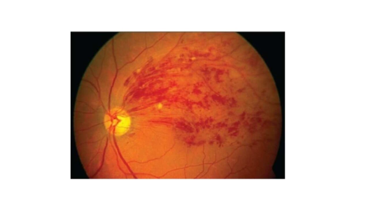
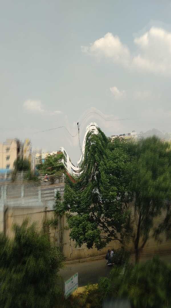
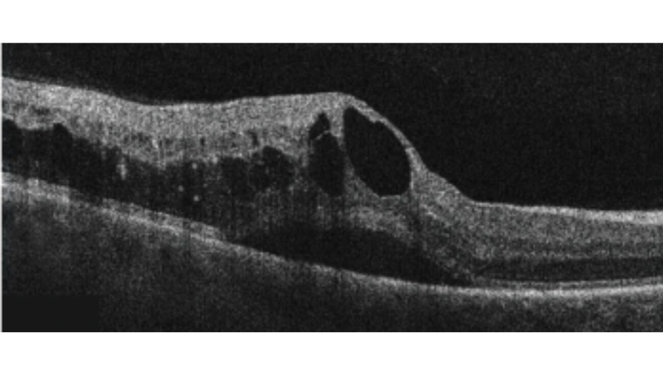
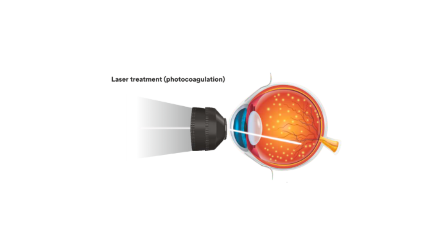
Retinal vein occlusion (RVO) is a blockage of the normal venous drainage from the retinal tissue commonly by a blood clot or another blood vessel. When the central vein is interrupted it is called the Central Retinal Vein Occlusion,(CRVO) and when a branch is occluded it’s called branch retinal vein occlusion. (BRAVO), Sometimes, the vein draining one half of the retina is blocked, it is called a Hemi Central Retinal Vein Occlusion,(Hemi-CRVO). It is frequently associated with macular edema (swelling of the central retinal layers) and vitreous hemorrhage ( bleeding in the vitreous cavity), which requires treatment with either intravitreal injections, retinal lasers, or surgery.

Branch retinal vein occlusion

Branch retinal vein occlusion

Central retinal vein occlusion

Central retinal vein occlusion

What are the risk factors of RVO?

Uncontrolled Hypertension is the most common risk factor followed by diabetes mellitus, dyslipidemia, glaucoma, use of oral contraceptive pills, smoking, and glaucoma. Branch retinal vein occlusion Central retinal vein occlusion Its usually seen in the elderly population, but uncommonly can be seen in young patients. Here the main causes can be due to hypercoagulable states, myeloproliferative disorders vasculitis, inflammatory disorders, infections, etc

What are the symptoms of RVO?

01

Blurring of central vision-Patients complains of painless, blurry vision making it difficult to read and write, sew and do near work.

02

Distorted central vision- this reduces the quality of vision, making it difficult to recognise faces and objects.

03

Black spots in the field of vision, also called as floaters or flashes of light is noted in the advanced stages. It occurs due to bleeding and retinal traction secondary to RVO complications.

intermediate stage of ARMD

What are the investigations advised for diagnosis of RVO ?

Retinal vein occlusion can be easily diagnosed by the retina specialist during clinical examination. Ancillary tests are done to detect complications like macular edema and neovascularisation.

OPTICAL COHERENCE TOMOGRAPHY (OCT) SCAN of the retina is advised to quantify
the amount of macular edema which will be useful while starting treatment.
OCT is a quick, non invasive test to image the retina. Here high resolution cross sectional images
(refer to image 1 and 2) are taken of the central part of retina called as macula where the CNVMs
are most commonly seen. After dilation of pupils, the patient is asked to sit upright on a chair in
front of the OCT machine and rest their chin on a chinrest and scans are taken.

OPTICAL COHERENCE TOMOGRAPHY (OCT) SCAN:

NORMAL OCT scan of the retina
normal otc scan in retina
FUNDUS PHOTOS:

Fundus photos are taken using a retinal camera to document the lesion and used to compare
during follow-ups.

FUNDUS FLUORESCEIN ANGIOGRAPHY:

This is an invasive technique to view retinal and choroidal blood vessels. A dye is injected via a
vein on the patient’s arm and a retinal camera is used to digitally record the images of the retinal
circulation.
This procedure is advised in selected cases of RVO to look for abnormal leaky vessels and
ischaemic areas in the retina.

FOR INVESTIGATING THE UNDERLYING CAUSE

The following tests may be advised -

Blood pressure, Erythrocyte sedimentation rate (ESR), Complete blood count (CBC), Random blood glucose, Lipid profile, Serum homocysteine, Renal profiles, Thyroid function tests, ECG, ECHO, carotid doppler. Additional tests like Thrombophilia screening, and autoantibodies, may be advised to rule out uncommon causes predisposing to RVO.

What are the treatment modalities for RVO?

Treatment of RVO is focused on vision-threatening complications such as macular edema and neovascularization. (abnormal vessel growth)

ANTI VEGF INJECTIONS:

ANTI VEGF INJECTIONS:

This is one of the main modality of treatment. It prevents vision loss, stabilizes vision, and improves vision in the presence of macular edema if administered at the right time. These include drugs like Bevacizumab, Ranibizumab, Aflibercept and Brolucizumab. They act by limiting the leakage from pathologic blood vessels and controlling disease activity. These injections are given as monthly and bimonthly doses depending on the molecule and disease severity and require regular follow-up as advised by the treating retina specialist.

Laser Treatment

RETINAL LASER

Two types of laser may be advised in RVO. In some cases, the retina specialist may plan macular laser which the leaking blood vessels and reduces macular edema. In other cases, a retinal photocoagulation laser is advised to treat ischaemic areas. This will reduce the disease activity and risk of bleeding in the future. Retinal laser is an OPD procedure that takes 10-15 minutes to be completed after dilatation.

ophthalmologist examining eyes of patient

INTRAVITREAL STEROIDS:

In certain case scenarios, the retina specialist may advise steroid injections in the form of dexamethasone implants based on the appearance of DME. The advantage of these implants is that it stays in the eye for a longer period resulting in less frequent injections. Some patients may develop high intraocular pressures and progression of cataracts post injection, hence proper case selection and follow up is highly important.

What is the prognosis of RVO and preventive measures?

With prompt diagnosis and early treatment, the prognosis is good, but since the disease takes a longer course,
regular follow-up is mandatory. Patients with ischemic CRVO are seen monthly for 6 months to detect the
onset of anterior segment neovascularization.
Preventive measures mainly concentrate on controlling systemic parameters like strict control of
Hypertension, Diabetes, and high cholesterol. Maintaining a healthy diet and regular exercise goes a long
way in controlling the above factors. Avoid smoking, regular retina checkups, at least once a year, and
follow whichever schedule is advised.

Author Details:

Dr. Sherina Thomas

Dr. Sherina Thomas

Medical retina specialist and Vitreo Retinal surgeon, MRCS Edin, FVRS.

vijaya nethralaya

© 2023 All Rights Reserved.

[html_block id="258"]
Sidebar