
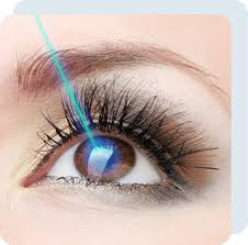
1. Laser Vision Correction (LASIK & SMILE):
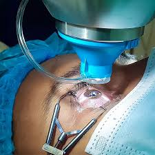
Correcting refractive errors like nearsightedness (myopia), farsightedness (hyperopia), and astigmatism to eliminate reliance on glasses or contact lenses is often achieved through an eye surgery procedure.
The core concept behind laser vision correction is altering how light bends as it enters the eye. By precisely reshaping the cornea (the clear front window of the eye), the surgeon enables light rays to focus directly on the retina (the light-sensitive layer at the back of the eye) instead of falling short or focusing behind it.
- LASIK: An ultra-fast laser (femtosecond laser) creates a microscopic, hinged flap on the surface of the cornea. The surgeon gently folds back the flap, and the excimer laser vaporizes micro-layers of tissue underneath to reshape the cornea. The surgeon then smooths the flap back down, where it acts as a natural bandage that heals without stitches.
- SMILE: Alternatively, this newer, flapless method allows a laser to carve a tiny, disc-shaped piece of tissue (a lenticule) inside the cornea, which the surgeon then removes through a microscopic keyhole incision.
2. Cataract surgery.
Replacing a cloudy natural lens with a clear artificial one to restore vibrant, sharp vision.
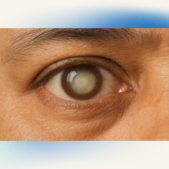
A cataract is a natural aging process in which proteins inside the eye’s crystalline lens clump together, causing the lens to become cloudy and making vision appear foggy or yellowed.
The standard procedure to correct this is phacoemulsification:
1. Micro-Incision: Step 1.
The surgeon makes a self-healing micro-incision (typically less than 2.5 mm) at the edge of the cornea.
2. Phacoemulsification: Step 2.
The surgeon introduces an ultrasonic probe vibrating at high frequencies into the eye, where it gently breaks the cloudy lens into microscopic pieces and then suctions them out.
3. IOL Implantation: Step 3.
Next, a flexible, folded intraocular lens (IOL) is carefully inserted through the same micro-incision. Once it enters the empty lens capsule, it gently unfolds into its permanent position.
3. Glaucoma & Vitreoretinal Surgeries:
Managing intraocular pressure (glaucoma) or repairing deep structural damage to the back of the eye (retinal tears/detachments).
Unlike elective vision correction, these specialized surgeries focus on preservation and stopping advanced diseases from causing permanent vision loss.
- Trabeculectomy (Glaucoma): When fluid cannot drain properly, intraocular pressure rises, damaging the optic nerve. A trabeculectomy creates a tiny, controlled drainage flap under the eyelid, allowing fluid to bypass the clogged internal drains and escape into a small reservoir, safely lowering eye pressure.
- Vitrectomy (Retina): To treat retinal detachments or severe bleeding from diabetic retinopathy, a surgeon removes the vitreous gel filling the center of the eye using a micro-suction tool. This provides clear access to fix or laser the retina, often filling the eye space with a temporary gas bubble or silicone oil to press the retina back into place while it heals.
The Patient Journey: What to Expect:
Before the Procedure
A comprehensive diagnostic workup maps the eye’s unique topography and measurements. On the day of surgery, we give you local numbing eye drops to ensure you feel absolutely no pain. In some instances, a mild oral or intravenous sedative is provided to help you stay calm and perfectly still.
During the Procedure
Most modern eye surgeries are remarkably brief, often lasting between 10 and 30 minutes per eye. You will remain awake, lying comfortably flat on your back, and will look at a fixed blinking target light to keep your eye aligned. A gentle spring-like instrument (a speculum) holds your eyelids open so you don’t have to worry about blinking.
Aftercare & Recovery:
Critical Recovery Rule: The first 24 to 48 hours are essential for proper structural healing. During this period, you must wear a protective shield while sleeping to prevent accidental rubbing, which could dislodge the flap or irritate fresh entry sites.
| Surgery Type | Typical Visual Improvement | Full Recovery Window | Key Restriction |
| LASIK / SMILE | Within 24 hours | 1 to 2 weeks | Absolutely no eye rubbing or swimming |
| Cataract | Within 2 to 3 days | 4 to 6 weeks | Avoid heavy lifting or bending forward |
| Vitrectomy | Gradual (weeks to months) | 4 to 8 weeks | Strict head positioning if a gas bubble is used |