

Eye health has become a major focus in modern preventive medicine, and diagnostic imaging plays a critical role in preserving vision. Among the most advanced technologies available today, the OCT vision exam stands out as one of the most precise and non-invasive methods for evaluating retinal and optic nerve health. Short for Optical Coherence Tomography, OCT has transformed the way eye specialists diagnose, monitor, and treat a wide range of ocular diseases. Instead of relying solely on external observation or conventional imaging, eye doctors can now examine microscopic layers of the retina with extraordinary detail.
Think of an OCT scan as the eye-care equivalent of an ultrasound—except instead of sound waves, it uses light waves to create high-resolution cross-sectional images. This allows ophthalmologists and optometrists to detect subtle structural changes long before noticeable symptoms appear. Doctors use OCT to screen for glaucoma, monitor diabetic eye disease, and evaluate age-related macular degeneration, making it a cornerstone of modern ophthalmic diagnostics.
With millions of OCT scans performed globally each year, this technology continues to improve diagnostic accuracy and patient outcomes. Understanding how an OCT eye exam works, why it matters, and what results can reveal can help patients make informed decisions about their visual health.
Understanding OCT Vision Exams
The OCT vision exam is designed to provide detailed images of the internal structures of the eye, particularly the retina, optic nerve, and macula. Unlike standard eye examinations that assess visual acuity or surface anatomy, OCT goes several layers deeper—literally. It captures microscopic images that allow specialists to identify swelling, thinning, fluid accumulation, and nerve damage.
One of the most impressive aspects of OCT technology is its ability to generate images in real time. The resulting scans look similar to topographical maps, where each retinal layer can be analyzed individually. This precision enables clinicians to identify diseases at their earliest stages, often before a patient notices changes in vision.
As preventive healthcare gains momentum, doctors increasingly include OCT in routine eye exams, especially for people over forty and those with diabetes, hypertension, or a family history of eye disease.
What Does OCT Stand For?
Optical Coherence Tomography combines three concepts:
- Optical refers to light-based imaging.
- Coherence involves measuring reflected light waves.
- Tomography means creating cross-sectional images.
In simple terms, OCT uses reflected light to create detailed, layered images of eye tissues. The technology measures how light bounces off different retinal structures and converts that information into digital images.
The result is incredibly precise imaging—often down to a few microns. For comparison, a human hair is roughly 70 microns thick, meaning OCT can detect changes much smaller than what the naked eye can observe.
How OCT Technology Works
OCT technology works by projecting low-coherence infrared light into the eye. As the light encounters different retinal layers, portions of it reflect back at varying speeds and intensities. The OCT machine collects this reflected light and uses sophisticated algorithms to reconstruct a cross-sectional image.
This process happens almost instantly, allowing clinicians to capture hundreds or even thousands of scans in seconds. Modern spectral-domain and swept-source OCT systems offer even higher resolution and deeper tissue penetration.
Because OCT uses light rather than radiation, it is considered extremely safe. There are no known harmful side effects, making it suitable for repeated monitoring over months or years.
The Evolution of OCT in Modern Ophthalmology
When OCT was first introduced in the early 1990s, it represented a breakthrough in retinal imaging. Before OCT, eye specialists relied heavily on fundus photography, fluorescein angiography, and manual examination methods.
Today’s OCT systems are exponentially faster and more accurate. High-definition imaging, three-dimensional reconstruction, and AI-assisted analysis have elevated OCT into one of the most trusted tools in ophthalmology.
According to industry reports, the global OCT market continues to expand due to increasing prevalence of chronic eye diseases and aging populations worldwide.
From Traditional Imaging to Advanced OCT
Traditional retinal imaging provided useful surface-level information, but often lacked the depth needed for early diagnosis. OCT changed that by allowing doctors to “slice” through retinal tissue virtually.
This advancement means specialists can now detect:
- Microscopic retinal swelling
- Optic nerve fiber thinning
- Macular fluid accumulation
- Early structural degeneration
These findings often appear months or years before symptoms become clinically obvious.
Current Global Adoption in Eye Care
OCT is now considered standard practice in:
| Medical Setting | OCT Usage |
|---|---|
| Ophthalmology Clinics | Disease diagnosis and monitoring |
| Optometry Practices | Preventive screening |
| Hospitals | Surgical planning |
| Research Institutions | Clinical studies |
| Diabetes Centers | Retinal screening |
Its widespread adoption reflects its reliability and clinical value.
Why Doctors Recommend an OCT Vision Exam
An OCT vision exam is often recommended when patients experience blurred vision, visual distortion, blind spots, or unexplained vision loss. However, many eye specialists also recommend OCT scans even when no symptoms are present.
Why? Because many serious eye diseases progress silently.
Conditions such as glaucoma may damage optic nerves for years before vision changes become noticeable. By the time symptoms appear, irreversible damage may already exist.
OCT allows clinicians to detect structural abnormalities early, significantly improving treatment outcomes.
Early Detection of Eye Diseases
Early diagnosis remains one of the strongest benefits of OCT. Structural changes in retinal layers often occur long before symptoms develop.
Examples include:
- Glaucoma nerve fiber loss
- Macular edema
- Retinal thinning
- Vascular abnormalities
Early intervention can preserve vision and reduce long-term complications.
Monitoring Chronic Eye Conditions
Patients with chronic eye diseases often undergo OCT scans regularly. Sequential scans help specialists compare changes over time.
This allows doctors to answer critical questions:
- Is treatment working?
- Is the disease progressing?
- Is surgery needed?
- Should medication be adjusted?
Such data-driven decision-making improves personalized care.
Conditions Diagnosed with OCT
Glaucoma Detection
Glaucoma is one of the leading causes of irreversible blindness worldwide. OCT helps measure the thickness of the retinal nerve fiber layer and optic nerve structures.
Even subtle thinning can indicate early glaucoma before visual field loss occurs.
Macular Degeneration Assessment
Age-related macular degeneration affects central vision and is common among older adults. OCT reveals drusen deposits, fluid accumulation, and tissue degeneration with remarkable precision.
This helps distinguish between dry and wet AMD.
Diabetic Retinopathy Evaluation
Diabetes can damage retinal blood vessels, causing leakage and swelling. OCT identifies:
- Macular edema
- Retinal thickening
- Fluid pockets
- Vascular complications
Early detection can prevent severe vision loss.
Retinal Detachment and Macular Hole
OCT provides detailed structural imaging that helps diagnose:
- Retinal tears
- Macular holes
- Vitreomacular traction
- Epiretinal membranes
This is essential for surgical planning.
What Happens During an OCT Vision Exam
An OCT exam is painless, fast, and non-invasive. Most patients find the procedure surprisingly simple.
Preparation Before the Test
Patients may be asked to remove glasses or contact lenses. In some cases, dilating drops may be used for clearer imaging.
No fasting or special preparation is typically required.
Step-by-Step Examination Process
- The patient sits in front of the OCT machine.
- Chin and forehead are positioned for stability.
- The patient focuses on an internal fixation target.
- The scanner captures multiple retinal images.
- Digital images are analyzed instantly.
The process requires minimal patient participation.
How Long the Scan Takes
Most OCT scans take 5 to 10 minutes, including setup and image review.
Actual image capture often takes less than one minute per eye.
Understanding OCT Scan Results
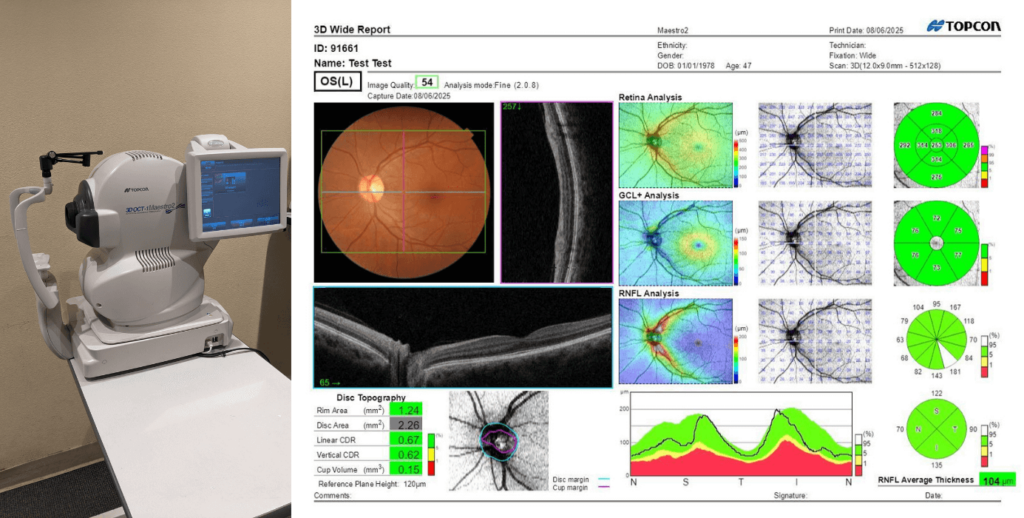
After the scan, specialists review retinal thickness maps, cross-sectional images, and comparative progression data.
Reading Retinal Layers
The retina contains multiple layers, each serving a unique function. OCT can distinguish these layers with micron-level accuracy.
Healthy layers appear smooth, organized, and symmetrical.
What Abnormal Findings May Mean
Abnormal findings may include:
| Finding | Possible Meaning |
|---|---|
| Retinal Thickening | Edema or inflammation |
| Thinning | Degeneration or glaucoma |
| Fluid Pockets | Wet macular degeneration |
| Tissue Separation | Retinal detachment |
| Irregular Surface | Epiretinal membrane |
Interpretation should always be performed by a qualified eye specialist.
Benefits and Limitations of OCT Vision Exams
Key Benefits for Patients and Specialists
The advantages of OCT are substantial:
- Non-invasive
- Radiation-free
- High-resolution imaging
- Fast results
- Early disease detection
- Long-term monitoring
For specialists, OCT provides objective measurements that improve treatment planning.
Possible Limitations and Challenges
Despite its strengths, OCT has limitations.
Poor image quality may occur due to:
- Cataracts
- Dry eyes
- Poor fixation
- Excessive blinking
Also, OCT provides structural—not always functional—information, meaning additional testing may still be required.
“OCT has fundamentally changed our ability to visualize living tissue at microscopic resolution.”
That statement captures exactly why OCT remains indispensable in modern eye care.
Conclusion
The OCT vision exam has become one of the most valuable diagnostic tools in ophthalmology, offering unmatched insight into retinal and optic nerve health. Its ability to detect microscopic structural changes before symptoms appear makes it essential for early diagnosis, preventive care, and ongoing disease management. Whether screening for glaucoma, monitoring diabetes-related complications, or assessing age-related macular degeneration, OCT empowers both clinicians and patients with precise, actionable information.
As eye care continues evolving with digital imaging and artificial intelligence, OCT remains at the center of modern vision preservation. For anyone concerned about long-term eye health, an OCT eye exam can be one of the smartest preventive decisions available.
Frequently Asked Questions
1. Is an OCT vision exam painful?
No, an OCT scan is completely painless and non-invasive. No needles, injections, or physical contact with the eye are involved.
2. How often should I get an OCT eye exam?
Frequency depends on age, risk factors, and medical history. High-risk patients may need scans every 6–12 months.
3. Does insurance cover OCT scans?
Many insurance providers cover medically necessary OCT exams, especially for glaucoma, diabetes, or retinal disease monitoring.
4. Can OCT detect glaucoma early?
Yes. OCT can detect optic nerve fiber thinning before noticeable vision loss occurs.
5. Do I need pupil dilation for an OCT scan?
Not always. Some scans can be performed without dilation, though dilation may improve image quality in certain cases.