

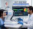
What Is an OCT Eye?
An OCT eye test (Optical Coherence Tomography) is a non-invasive imaging scan that uses light waves to capture high-resolution cross-sectional images of the retina, allowing doctors to detect early signs of eye diseases like glaucoma, macular degeneration, and diabetic retinopathy—often before symptoms appear.
Understanding Optical Coherence Tomography:
If you’ve ever wondered how eye doctors can “see” beneath the surface of your retina without touching your eye, OCT is the answer. Think of it like an ultrasound—but instead of sound waves, it uses light waves to create incredibly detailed images of the retina’s internal structure. This technology has completely changed how eye conditions are diagnosed, shifting from guesswork to precision imaging.
OCT scans work at a microscopic level, producing images that are so detailed they can measure retinal thickness down to a few microns. That’s thinner than a human hair. This level of precision allows ophthalmologists to detect diseases years before vision loss occurs, which is a game changer for prevention.
What makes OCT especially powerful is its ability to provide a layer-by-layer view of the retina. Instead of just seeing the surface, doctors can analyze each structural layer individually—almost like peeling back the pages of a book to understand the whole story.
How OCT Differs from Traditional Eye Exams:
Traditional eye exams—like vision tests or slit-lamp exams—are essential, but they mostly focus on what’s visible on the surface. OCT, on the other hand, dives deeper. It reveals structural changes that are invisible during routine checkups.
For example, you might have perfect vision on a standard eye chart, yet OCT could still detect early signs of glaucoma or macular degeneration. That’s because these diseases often begin silently, without noticeable symptoms.
Another key difference lies in objectivity. Traditional exams rely heavily on the doctor’s observation, while OCT provides quantifiable data—measurable thickness maps, nerve fiber analysis, and progression tracking over time. This makes it easier to monitor disease progression and adjust treatment plans accurately.
Patients often ask, “Do I really need an OCT if my vision is fine?” The honest answer: yes, in many cases. Especially if you’re at risk, OCT acts like an early warning system. It’s the difference between reacting to vision loss and preventing it altogether.
How OCT Technology Works
OCT technology uses low-coherence interferometry with reflected light waves to generate detailed 3D images of the retina, enabling precise measurement of tissue thickness and structure in real time without any physical contact or radiation exposure.
The Role of Light and Interferometry
At its core, OCT relies on a fascinating principle called interferometry. Without getting overly technical, it works by splitting a beam of light into two paths—one directed into the eye and the other used as a reference. When these beams reflect back, they create an interference pattern that is analyzed to form detailed images.
It sounds complex, but the result is simple: a real-time map of your retina’s internal layers. The machine processes thousands of light reflections per second, building a cross-sectional image that looks almost like a slice through the eye.
One of the biggest advantages here is safety. Unlike X-rays or CT scans, OCT uses non-ionizing light, meaning there’s no radiation exposure. This makes it safe for repeated use, even in children or patients requiring frequent monitoring.
Another underrated benefit is speed. Most OCT scans take less than 5 minutes, and results are available instantly. This allows doctors to make decisions on the spot, rather than waiting days for lab reports.
Spectral Domain vs Swept Source OCT
Key Differences in Accuracy and Speed
Not all OCT machines are created equal. In 2026, the two most widely used types are Spectral Domain OCT (SD-OCT) and Swept Source OCT (SS-OCT)—and the difference between them can directly impact diagnostic accuracy.
| Feature | Spectral Domain OCT | Swept Source OCT |
|---|---|---|
| Light Source | Broadband light | Tunable laser |
| Speed | Moderate | Ultra-fast |
| Depth Penetration | Limited | Deeper imaging |
| Image Clarity | High | Very high |
| Cost | More affordable | More expensive |
SD-OCT has been the industry standard for years and is still widely used in clinics across Bengaluru. It offers excellent resolution and is sufficient for most routine diagnoses.
SS-OCT, however, is the newer, more advanced option. It penetrates deeper into the eye, allowing doctors to visualize structures like the choroid more clearly. This is especially useful for complex cases, such as high myopia or advanced retinal diseases.
The trade-off? Cost. Clinics using SS-OCT often charge higher fees, but they also provide more comprehensive insights. If you’re dealing with a complicated condition, opting for SS-OCT could be worth it.
The 10 Retinal Layers Explained (Direct Answer Summary):
The retina consists of 10 distinct layers, each playing a critical role in vision; OCT imaging allows clinicians to analyze these layers individually, helping detect early structural abnormalities associated with diseases like glaucoma, macular degeneration, and diabetic retinopathy.
Why Retinal Layer Analysis Matters
Imagine trying to diagnose a structural problem in a building without seeing its internal framework. That’s what eye care was like before OCT. Now, doctors can examine each of the 10 retinal layers separately, identifying exactly where things go wrong.
These layers include:
- Retinal Nerve Fiber Layer (RNFL)
- Ganglion Cell Layer
- Inner Plexiform Layer
- Inner Nuclear Layer
- Outer Plexiform Layer
- Outer Nuclear Layer
- Photoreceptor Layer
- Retinal Pigment Epithelium (RPE)
Each layer has a specific function, from transmitting visual signals to absorbing excess light. When a disease begins, it often affects one particular layer first. OCT helps pinpoint this early damage.
For example, glaucoma typically starts with thinning of the RNFL, while macular degeneration affects the RPE and photoreceptor layers. By catching these subtle changes early, doctors can intervene before irreversible damage occurs.
Diseases Detected Through Layer Changes
OCT isn’t just about pretty images—it’s about actionable insights. Changes in retinal layers can signal a wide range of conditions, sometimes even before symptoms appear.
For instance, studies published on PubMed have shown that OCT can detect diabetic retinal changes up to 5 years before vision loss begins. That’s a huge window for preventive care.
Some key conditions identified through OCT include:
- Glaucoma (nerve fiber thinning)
- Age-related macular degeneration (AMD) (drusen deposits)
- Diabetic retinopathy (fluid accumulation)
- Macular edema
- Retinal detachment
What’s fascinating is how OCT allows doctors to track these conditions over time. It’s not just a snapshot—it’s a timeline. By comparing scans, clinicians can see whether a disease is progressing, stabilizing, or improving with treatment.
Key Benefits of OCT Eye Tests
OCT eye tests provide early, highly accurate detection of retinal and optic nerve diseases, enable real-time monitoring of disease progression, and support personalized treatment decisions—all through a fast, non-invasive, and completely painless scanning process.
Early Detection of Eye Diseases
One of the biggest advantages of an OCT eye test is its ability to detect diseases long before you notice anything wrong with your vision. That’s not just helpful—it’s often the difference between preserving your sight and facing irreversible damage. Many serious eye conditions, including glaucoma, diabetic retinopathy, and macular degeneration, develop silently. By the time symptoms like blurred vision appear, the disease may already be in an advanced stage.
OCT flips that timeline completely. It identifies microscopic structural changes in the retina, sometimes years before symptoms begin. Think of it like catching cracks in a dam before it bursts—early detection gives doctors time to act, adjust, and prevent disaster.
The early intervention can reduce the risk of severe vision loss in glaucoma patients by up to 50%. OCT plays a central role in that early intervention. It measures the thickness of the retinal nerve fiber layer (RNFL), which is one of the first areas affected in glaucoma. Even slight thinning can trigger further investigation and early treatment.
What makes this even more powerful is the objectivity of OCT data. Instead of relying on subjective symptoms or visual acuity tests, doctors get hard numerical evidence. This allows for more confident diagnoses and reduces the chances of missing subtle warning signs.
Monitoring Chronic Eye Conditions
If early detection is the first win, continuous monitoring is the long-term strategy—and OCT excels at that too. For patients already diagnosed with chronic conditions, OCT acts like a progress tracker, showing exactly how the disease is evolving over time.
Take diabetic retinopathy as an example. Patients with diabetes are at constant risk of developing retinal swelling or fluid accumulation. OCT can detect even tiny amounts of fluid in the retina, helping doctors decide when to start or adjust treatments like anti-VEGF injections. Without OCT, these decisions would rely heavily on symptoms, which can be misleading or delayed.
The same applies to macular degeneration, where OCT helps monitor the buildup of drusen or fluid under the retina. By comparing scans over weeks or months, doctors can see whether a treatment is working or if a different approach is needed.
Patients often find reassurance in this process. Instead of wondering whether their condition is getting worse, they can see the data visually. It turns an abstract health concern into something concrete and manageable.
OCT Applications in 2026
In 2026, OCT technology extends beyond ophthalmology into neurology, pediatrics, and preventive healthcare, enabling early detection of systemic diseases, brain-related conditions, and childhood vision disorders through advanced retinal imaging and AI-assisted diagnostics.
Neurology and Brain Health Insights
Here’s where things get really interesting—OCT is no longer just an eye test. Researchers and clinicians are increasingly using it as a window into the brain. Why? Because the retina is technically part of the central nervous system, meaning changes in the brain can sometimes show up in the eyes.
Recent studies cited in journals indexed on PubMed have demonstrated correlations between retinal nerve fiber thinning and neurodegenerative diseases like Alzheimer’s and Parkinson’s. In simple terms, OCT might help detect early signs of these conditions—sometimes even before cognitive symptoms appear.
This opens up a completely new frontier for preventive medicine. Imagine going for a routine eye scan and getting insights into your neurological health. It’s not science fiction—it’s already happening in advanced clinics.
AI integration is pushing this even further. Modern OCT systems can analyze patterns across thousands of scans, identifying subtle anomalies that might escape the human eye. These systems act like a second pair of expert eyes, enhancing diagnostic accuracy.
While OCT is not yet a standalone diagnostic tool for neurological diseases, it’s becoming an important screening and research instrument. As technology evolves, its role in brain health is expected to grow significantly.
Pediatric and Preventive Eye Care
Children’s eye care has traditionally been challenging. Kids may struggle to communicate vision problems, and standard tests often rely on subjective responses. OCT changes that dynamic completely by providing objective, image-based diagnostics.
In pediatric care, OCT is used to detect conditions like:
- Amblyopia (lazy eye)
- Congenital retinal disorders
- Optic nerve abnormalities
Because the test is quick and non-invasive, it’s ideal for children—even those who are anxious or uncooperative. Many clinics now use child-friendly OCT machines with faster scan times and visual aids to keep kids engaged.
Preventive care is another area where OCT shines. Instead of waiting for symptoms to appear, doctors can establish a baseline scan early in life and monitor changes over time. This is particularly useful for children with a family history of eye diseases.
In 2026, preventive healthcare is becoming more proactive than reactive. OCT fits perfectly into this model, offering a way to detect risks early and take action before problems escalate.
OCT vs Other Eye Tests
OCT differs from traditional eye tests by providing cross-sectional, high-resolution images of retinal structures, whereas tests like fundus photography and dilated exams offer surface-level views, making OCT significantly more effective for early detection and disease monitoring.
OCT vs Fundus Photography
At first glance, fundus photography and OCT might seem similar—they both involve imaging the retina. But the difference lies in depth and detail. Fundus photography captures a 2D image of the retinal surface, much like taking a photograph of a landscape.
OCT, on the other hand, is more like slicing that landscape into layers and examining each one individually. It provides cross-sectional images, revealing what’s happening beneath the surface.
Fundus photography is excellent for documenting visible changes like bleeding, lesions, or pigment alterations. It’s widely used for screening and record-keeping. However, it cannot detect subtle structural changes within the retinal layers.
That’s where OCT takes the lead. For conditions like glaucoma or early macular degeneration, OCT can identify problems long before they become visible in fundus images.
In many clinics, the two tests are used together rather than as alternatives. Fundus photography provides a broad overview, while OCT offers a detailed internal analysis. Think of it as using both a map and a microscope—you get the full picture.
OCT vs Dilated Eye Exams
A dilated eye exam is another common diagnostic tool, where eye drops are used to widen the pupil, allowing the doctor to examine the retina more thoroughly. While this method is effective, it still relies heavily on visual inspection.
OCT eliminates that limitation by providing quantitative data and precise measurements. Instead of just observing the retina, doctors can measure thickness, detect fluid, and track changes over time.
There’s also a comfort factor to consider. Dilated exams can cause temporary side effects like light sensitivity and blurred vision, which can last for several hours. OCT, in contrast, has no downtime. You can walk in, get scanned, and resume your day immediately.
That said, dilated exams are still important. They allow doctors to assess areas that OCT might not fully capture, such as the peripheral retina. The best approach often combines both methods for a comprehensive evaluation.
Cost of OCT Eye Tests in Bengaluru (2026)
Average Price Breakdown Across Clinics
When it comes to healthcare decisions, cost is always part of the equation—and OCT testing in Bengaluru offers a wide range of pricing options depending on the clinic, technology, and expertise involved.
On average in 2026, the cost of an OCT eye test in Bengaluru falls within this range:
| Clinic Type | Price Range (INR) |
|---|---|
| Small Eye Clinics | ₹1,500 – ₹2,500 |
| Mid-Range Hospitals | ₹2,500 – ₹4,000 |
| Premium Eye Centers | ₹4,000 – ₹7,000 |
Smaller clinics often use Spectral Domain OCT, which keeps costs lower while still providing reliable results. Mid-range hospitals may include OCT as part of a broader diagnostic package, offering better value for patients needing multiple tests.
Premium centers, on the other hand, typically invest in Swept Source OCT and AI-assisted diagnostics. While the cost is higher, these facilities often provide more comprehensive analysis, shorter wait times, and access to experienced specialists.
It’s also worth noting that some clinics offer package deals, especially for diabetic or glaucoma patients who require regular monitoring. These packages can significantly reduce the per-scan cost over time.
Factors Affecting OCT Test Pricing
The price of an OCT test isn’t random—it’s influenced by several key factors. Understanding these can help you make a more informed decision.
First, technology matters. Clinics using advanced SS-OCT machines generally charge more due to higher equipment costs and maintenance. Second, the expertise of the doctor plays a role. Highly experienced ophthalmologists or retina specialists may charge a premium for their interpretation of the scan.
Location is another factor. Clinics in central or high-demand areas of Bengaluru tend to have higher pricing compared to those in suburban regions.
Finally, additional services—such as detailed reports, follow-up consultations, or combined diagnostic tests—can increase the overall cost. While it might be tempting to go for the cheapest option, it’s important to consider the quality and accuracy of the diagnosis.
Is an OCT Eye Test Safe and Painful?
An OCT eye test is about as straightforward and comfortable as medical diagnostics get. If you’re picturing needles, contact lenses, or anything invasive, you can relax—none of that is involved. The test is completely non-contact, meaning the machine doesn’t touch your eye at all.
You’ll typically be asked to sit in front of the device, rest your chin on a support, and focus on a small light. The scan itself takes just a few seconds per eye. There’s no discomfort, no pressure, and no recovery time needed afterward.
Safety is another strong point. OCT uses low-intensity light waves, not radiation, making it safe for repeated use. This is especially important for patients who need frequent monitoring, such as those with chronic eye conditions.
Even sensitive groups—like children, older patients, or individuals with underlying health conditions—can safely undergo OCT scans. There are no known long-term side effects, and the procedure has been widely validated by global health organizations, including the AAO.
If you’ve been avoiding an eye test due to fear or uncertainty, OCT is one of the easiest entry points into modern eye care.
When Should You Get an OCT Scan?
Timing matters more than most people realize. Many assume that eye tests are only necessary when vision problems appear, but with OCT, the goal is to stay ahead of symptoms, not react to them.
You should consider getting an OCT scan if you fall into any of these categories:
- Over the age of 40
- Diabetic or at risk of diabetes
- Family history of glaucoma or macular degeneration
- Experiencing blurred or distorted vision
- Already diagnosed with an eye condition
Even if none of these apply, a baseline OCT scan can be incredibly useful. It provides a reference point for future comparisons, making it easier to detect subtle changes over time.
Doctors often recommend annual or biennial OCT scans for high-risk individuals. For others, it may be part of a routine comprehensive eye exam every few years.
The key idea here is simple: don’t wait for symptoms. By the time vision changes become noticeable, some damage may already be irreversible. OCT gives you a chance to act early—and that’s invaluable.
FAQs
1. Is an OCT eye test painful?
Not at all. It’s completely painless and non-invasive. Most patients describe it as similar to having a photo taken of their eye.
2. How long does an OCT scan take?
The entire process usually takes 5–10 minutes, including setup and image capture.
3. Do I need dilation for an OCT test?
In many cases, dilation isn’t required. However, your doctor may recommend it for better imaging in certain situations.
4. How often should I get an OCT scan?
It depends on your risk factors. High-risk individuals may need annual scans, while others can do it less frequently.
5. Can OCT detect all eye diseases?
While OCT is highly advanced, it’s usually used alongside other tests for a comprehensive diagnosis.
Conclusion:
OCT eye testing has evolved from a specialized diagnostic tool into a cornerstone of modern healthcare. It offers a rare combination of precision, safety, and accessibility, making it one of the most valuable advancements in ophthalmology.
From detecting silent diseases to monitoring chronic conditions and even offering insights into brain health, OCT is redefining what an eye exam can achieve. In a fast-growing medical hub like Bengaluru, access to this technology is better than ever, with options available for every budget and need.
If there’s one takeaway, it’s this: clarity isn’t just about vision—it’s about understanding what’s happening beneath the surface. And OCT delivers exactly that.