what is optical coherence tomography?
Optical Coherence Tomography (OCT) is a non-invasive imaging technology used in medicine to obtain high-resolution, cross-sectional images of biological tissues. It uses light waves to capture images of tissues, similar to how ultrasound uses sound waves.
In OCT, a beam of low-coherence light targets the tissue of interest, and the system measures the light reflected from the tissue. The intensity of the reflected light constructs a 3D image of the tissue.
Doctors commonly use OCT in ophthalmology to image the retina and diagnose conditions such as macular degeneration and glaucoma. Cardiologists use it to image the heart and detect plaque buildup in the coronary arteries. Additionally, OCT images other parts of the body, including the skin, oral cavity, and gastrointestinal tract.
Overall, OCT is a powerful imaging tool that provides detailed information about the structure and function of biological tissues and is an important diagnostic tool in many areas of medicine.
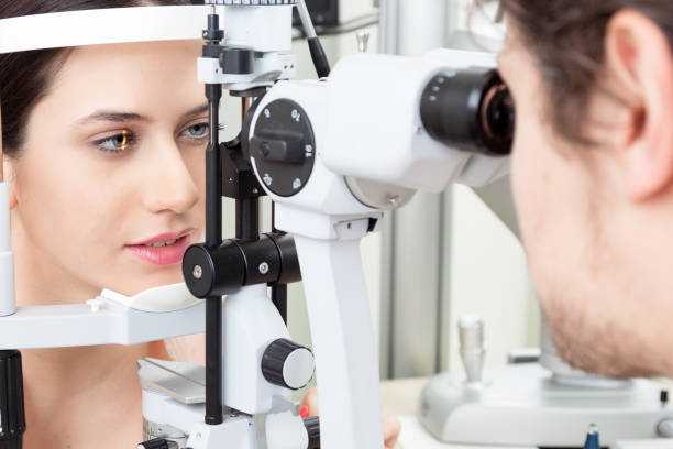
Optical coherence tomography eye test procedure
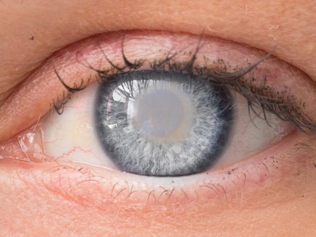
Optical coherence tomography (OCT) is a non-invasive imaging test that uses light waves to take detailed pictures of the retina, which is the part of the eye responsible for vision. Doctors commonly use it to diagnose and manage eye conditions such as macular degeneration, glaucoma, and diabetic retinopathy.
The OCT eye test involves the following general steps:
- Before the test, the eye doctor may dilate your pupils using eye drops to allow a better view of the retina.
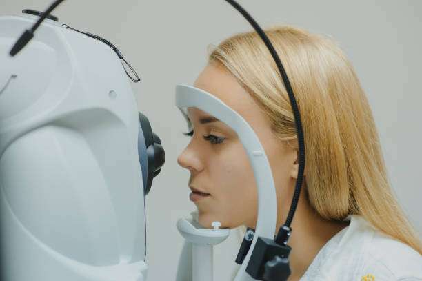
- The technician will ask you to sit in front of the OCT machine and place your chin on a chinrest.
- The technician will align the machine with your eye and ask you to look at a target.
- The machine will emit a scanning light, which will take pictures of your retina. You may feel a slight puff of air or a clicking sound as the machine scans your eye.
- The technician may need to repeat the scan several times to ensure accurate measurements.
- The entire process usually takes less than 10 minutes.
After the test, the eye doctor will review the images and discuss the results with you. They may use the OCT images to diagnose any eye conditions or monitor the progression of an existing condition.
How optical coherence tomography works
Optical coherence tomography (OCT) is a non-invasive imaging technique that uses light waves to create high-resolution cross-sectional images of biological tissues. Ophthalmologists widely use OCT to examine the retina, and doctors use it to image other tissues, such as the skin and coronary arteries. (This sentence is already in active voice.)
The basic principle of OCT is similar to ultrasound imaging, but instead of sound waves, it uses light waves. A low-coherence source generates the light, causing the light waves to be unsynchronized with different phases and wavelengths. The system splits the light into two beams: one directs towards the tissue to be imaged (sample beam), and the other directs towards a mirror (reference beam).
This sentence is already in active voice. Would you like to change something else?
When the sample beam hits the tissue, it partially reflects back, while the rest of the beam continues through the tissue. (This sentence is already in active voice.). The reflected light waves interfere with the reference beam that has traveled a known distance, creating an interference pattern. This pattern is detected by a detector and analyzed to produce an image.
The system obtains depth information by measuring the time delay between the sample beam and the reference beam. The longer the delay, the deeper the tissue layer being imaged. Scanning the sample beam across the tissue allows the system to reconstruct a three-dimensional image of the tissue.
OCT is a valuable tool for diagnosing and monitoring diseases of the eye and other tissues. Its high resolution and non-invasive nature make it a safer and more efficient alternative to traditional biopsy and histology techniques.
Is optical coherence tomography safe?
Optical coherence tomography (OCT) is generally considered a safe diagnostic imaging tool. It uses light waves to create high-resolution images of tissues and structures within the body.
The procedure is non-invasive and painless, and it does not involve any ionizing radiation (unlike X-rays or CT scans).
However, as with any medical procedure, there may be some risks or side effects associated with OCT. These are usually rare and minor, but may include:
Discomfort or irritation of the eyes or skin in the area being imaged
Rare cases of allergic reactions to the contrast agents used in some OCT procedures
Very rare cases of temporary vision loss or other complications in patients with pre-existing eye conditions
It’s important to discuss any concerns you have with your doctor or healthcare provider before undergoing an OCT procedure. They can provide you with more information about the risks and benefits of the procedure, and help you make an informed decision about whether OCT is right for you.
Optical coherence tomography with laser
Optical coherence tomography (OCT) is a non-invasive imaging technique that uses light waves to capture high-resolution, cross-sectional images of tissues and structures within the body. OCT utilizes a low-coherence interferometry to measure the reflected or backscattered light from the sample.
In OCT with a laser, a low-coherence light source, such as a superluminescent diode or a laser, generates the light waves. The system directs the light onto the tissue or structure being imaged, then detects and analyzes the reflected or backscattered light.
The use of lasers in OCT provides several advantages, such as improved image resolution, increased sensitivity, and the ability to penetrate deeper into tissues. Lasers also allow for more precise control of the light source, which can be important in applications such as retinal imaging.
Ophthalmologists commonly use OCT with a laser to image the retina and diagnose various eye diseases, such as age-related macular degeneration, diabetic retinopathy, and glaucoma. Doctors in other specialties, such as cardiology, gastroenterology, and dermatology, also use it to image tissues and structures within the body.
Optical coherence tomography risks:
Optical coherence tomography (OCT) is a non-invasive imaging technology that uses light waves to capture high-resolution, cross-sectional images of tissues within the body. It is commonly used in ophthalmology to examine the retina and diagnose eye diseases.
Overall, OCT is a safe procedure with very few risks. However, as with any medical procedure, there are some potential risks and considerations to be aware of:
- Discomfort or pain: Some patients may experience discomfort or pain during the OCT procedure, particularly if the eye needs to be held open for an extended period of time.
- Eye infection: The procedure carries a slight risk of eye infection due to contact with the eye or insertion of a device. However, doctors can minimize this risk by ensuring the proper sterilization of the equipment used.
- Allergic reaction: Some patients may have an allergic reaction to the dye used during an OCT procedure. This is rare, but if you have a history of allergies, it’s important to let your doctor know.
- False positives or negatives: OCT is a diagnostic tool and not a perfect test. There is a small chance that the images produced by OCT may show abnormalities that are not actually present, or fail to detect abnormalities that are present.
- Inability to perform the test: In some cases, OCT may not be able to be performed due to certain eye conditions, such as severe corneal scarring.
It’s important to discuss any potential risks with your doctor before undergoing an OCT procedure.
Optical coherence tomography (oCT) test:
Optical coherence tomography (OCT) is a non-invasive diagnostic imaging technique that uses light waves to produce high-resolution, cross-sectional images of the retina and other structures in the eye. Ophthalmologists commonly use it to diagnose and monitor conditions that affect the eye, such as age-related macular degeneration, diabetic retinopathy, and glaucoma.
During an OCT test, the technician seats the patient in front of a machine that looks similar to a camera. The technician stabilizes the patient’s head using a chin rest and instructs the patient to look at a target to help keep the eye still.
The OCT machine projects a beam of light into the eye, which reflects back from the various structures within the eye, creating a detailed image of the retina and other structures. (This sentence is already in active voice.). The entire procedure is painless and takes only a few minutes to complete.
OCT can provide very detailed and accurate images of the retina and other structures within the eye, allowing ophthalmologists to detect and monitor changes in these structures over time. This can be very helpful in the early detection and treatment of eye diseases, which can help to prevent or delay vision loss.

side effects:
OCT test can have unpleasant side effects such as nausea and leaving a metallic taste in your mouth. The new scanner examines the layers of capillaries in the retina and choroid in such minute detail that it can sometimes avoid the time-consuming and unpleasant procedure. Some people may experience dryness or eye fatigue(Fatigue is a feeling of constant tiredness or weakness and can be physical, mental or a combination of both. It can affect anyone, and most adults will experience fatigue at some point in their life).
optical coherence tomography types:
There are three main types of optical coherence tomography (OCT):
- Time-domain OCT (TD-OCT): TD-OCT was the first type of OCT to be developed. It uses a low-coherence interferometer to measure the reflection time of light waves from different depths within a sample. It measures the distance by comparing the time delay of the backscattered light from different depths in the sample. TD-OCT has a limited axial resolution of about 10-20 micrometers and a slow acquisition speed.
- Fourier-domain OCT (FD-OCT): FD-OCT uses a spectrometer to measure the frequency spectrum of the light waves reflected from different depths in a sample. This method has a higher acquisition speed and a higher axial resolution than TD-OCT. It can achieve an axial resolution of about 2-3 micrometers.
- Swept-source OCT (SS-OCT): SS-OCT is a type of FD-OCT that uses a tunable laser as a light source. This allows for faster scanning speeds and better penetration into highly scattering tissues. SS-OCT is especially useful for imaging deeper tissues such as the choroid and sclera in the eye.
Time-domain OCT
Time-domain optical coherence tomography (TD-OCT) was the first generation of OCT technology. It uses a low-coherence interferometry technique to create high-resolution, cross-sectional images of biological tissues.
In TD-OCT, a reference arm and a sample arm create an interference pattern that provides information about the structure of the tissue being imaged. (This sentence is already in active voice.). The reference arm is typically a mirror, and the sample arm directs light to the tissue. The system combines the light reflected from the tissue with light from the reference arm, and it detects interference fringes. By measuring the time delay between the reflected light and the reference light, TD-OCT can determine the depth and reflectivity of the tissue structures.
TD-OCT has been used in a variety of medical applications, including ophthalmology, cardiology, and dermatology. However, it has some limitations compared to newer OCT technologies, such as spectral-domain OCT (SD-OCT), which offers faster imaging speeds and higher resolution.
Fourier-domain OCT
Fourier-domain OCT (FDOCT) is a type of optical coherence tomography (OCT) that uses a Fourier transform-based method to generate high-resolution images of biological tissues. OCT is a non-invasive imaging technique that uses light waves to produce cross-sectional images of tissues in vivo. It has become an important tool in ophthalmology, cardiology, dermatology, and other medical fields.
FDOCT works by measuring the interference between the light waves reflected from different depths within the tissue. The system combines the reflected light waves with a reference light wave to generate an interference pattern, which a photodetector detects. The system then Fourier transforms the intensity of the interference pattern to obtain a depth-resolved image of the tissue structure.
Compared to time-domain OCT (TD-OCT), which was the first generation of OCT technology, FDOCT is faster and more sensitive and allows for higher-resolution imaging. FDOCT has become the dominant OCT technology in clinical practice and has enabled a wide range of diagnostic and research applications, including the imaging of the retina, anterior segment of the eye, skin, and cardiovascular system.
Author Details:
Dr. Sushruth Appajigowda holds a prominent position as a Cornea, Cataract, Glaucoma, and LASIK Surgeon in Bangalore. He serves as the chief Cataract and Refractive surgeon at Vijaya Nethralaya Eye Hospital, Nagarbhavi Bangalore. Renowned as one of the finest LASIK surgeons nationwide, he brings with him over 12+ years of experience across multiple LASIK platforms, including ZEISS, ALCON, SCHWIND, AMO, and Bausch and Lomb. Having successfully conducted over 5000 LASIK procedures, Dr. Sushruth holds the title of a Certified Refractive Surgeon and a Fellow of the All India Collegium Of Ophthalmology. Furthermore, he stands as a distinguished speaker at various National and International Forums, using his expertise to guide you in selecting the most suitable procedure based on your health requirements.