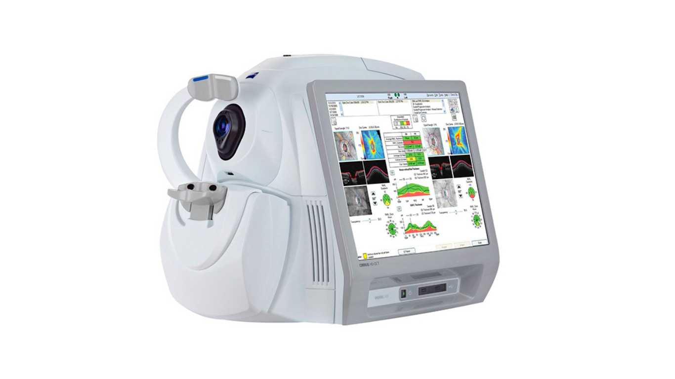
In the realm of ophthalmology, the OCT (Optical Coherence Tomography) test has emerged as a revolutionary tool in the diagnosis and management of various eye conditions, including glaucoma. Leveraging its non-invasive and highly detailed imaging capabilities, the OCT test plays a crucial role in the early detection and monitoring of glaucoma, a serious eye disease that can lead to irreversible vision loss if not properly managed. In this article, we’ll delve into the intricacies of the OCT scanning for glaucoma, its significance, and how it contributes to the overall eye health of patients.
Introduction:
Glaucoma, often referred to as the “silent thief of sight,” is a group of eye diseases that damage the optic nerve, leading to gradual vision loss. Consequently, it is imperative to detect and manage glaucoma at an early stage to prevent permanent damage. This is precisely where the OCT test steps in as a game-changing diagnostic tool.
Understanding Glaucoma: A Silent Threat to Vision:
Glaucoma is notorious for its asymptomatic nature during the early stages. Consequently, patients may not experience noticeable symptoms until significant vision loss occurs. This highlights the urgency of regular eye examinations, including comprehensive glaucoma evaluations.
What is OCT scanning for glaucoma ?
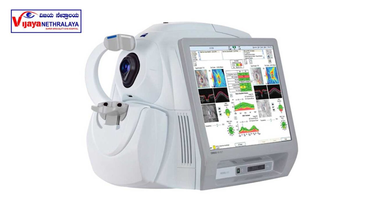
Optical Coherence Tomography (OCT) is a non-invasive imaging technique that furnishes high-resolution cross-sectional images of ocular structures.
The Role of OCT in Glaucoma Diagnosis:
OCT’s precision in capturing detailed images allows ophthalmologists to visualize subtle structural changes caused by glaucoma. This early detection aids in devising appropriate treatment plans and preventing irreversible vision loss.
Early Detection: A Game Changer in Glaucoma Management
Early detection of glaucoma significantly enhances the effectiveness of treatment strategies. As a result, timely identification allows for more successful implementation of therapeutic approaches. OCT aids in identifying even minor structural changes, enabling timely interventions to control the disease’s progression.
How Does the OCT Test Work?
The OCT test involves directing a beam of light into the eye. As a result, the reflected light produces detailed, cross-sectional images of ocular structures. These images, in turn, greatly assist ophthalmologists in identifying abnormalities and effectively tracking the progression of diseases.
Types of OCT Imaging in Glaucoma Assessment
OCT offers diverse imaging modes, including time-domain OCT (TD-OCT) and spectral-domain OCT (SD-OCT), each with unique advantages. SD-OCT, for instance, provides faster scans and higher-resolution images. Moreover, this technology improves diagnostic accuracy and facilitates more precise assessments in various medical applications.
Advantages of OCT over Traditional Diagnostic Methods
Compared to traditional methods, OCT is non-invasive and delivers real-time, high-resolution images. This eliminates the need for invasive procedures and enhances patient comfort.
OCT Angiography: Unveiling Microvascular Changes
OCT angiography, being an extension of OCT, significantly enhances the ability to visualize retinal blood vessels. This, in turn, greatly aids in comprehending microvascular changes linked to glaucoma. Consequently, it plays a pivotal role in facilitating early detection of this condition.
Tracking Progression: Monitoring with OCT
OCT’s remarkable ability to detect subtle changes over time renders it an invaluable tool for effectively monitoring the progression of glaucoma. This, in turn, empowers ophthalmologists with the capability to make necessary adjustments to treatment plans.
Ensuring Patient Comfort During OCT Testing:
OCT testing offers a quick, painless, and non-invasive procedure for patients. During the test, individuals only need to focus on a designated target as the device captures images. This streamlined process ensures patient comfort and cooperation, enhancing the overall reliability of the results. This seamless process ensures a comfortable and hassle-free experience for patients undergoing the procedure.
The Expertise of Ophthalmologists in Interpreting OCT Results
Interpreting OCT results necessitates specialized training, owing to the complexity of the images generated. Consequently, ophthalmologists meticulously analyze the captured images, leveraging their expertise to ensure precise diagnoses and the formulation of highly effective treatment strategies.
Future Potential: Research and Advancements in OCT Technology
OCT technology is in a constant state of evolution, and ongoing research plays a pivotal role in augmenting its capabilities. Innovations such as enhanced depth imaging (EDI-OCT) are paving the way for even more comprehensive imaging of ocular structures. This promises to provide healthcare professionals with increasingly detailed insights for accurate diagnoses and effective treatment planning.
OCT Beyond Glaucoma: A Versatile Diagnostic Tool
Playing a pivotal role in the realm of glaucoma management, OCT also exhibits applications that extend beyond the field of ophthalmology. Moreover, this versatile technology finds utility in diverse disciplines such as neurology, dermatology, and cardiology. This underscores its wide-ranging capabilities and potential impact across various medical specialties.
Author Details:
Dr. Sushruth Appajigowda holds a prominent position as a Cornea, Cataract, Glaucoma, and LASIK Surgeon in Bangalore. He serves as the chief Cataract and Refractive surgeon at Vijaya Nethralaya Eye Hospital, Nagarbhavi Bangalore. Renowned as one of the finest LASIK surgeons nationwide, he brings with him over 12+ years of experience across multiple LASIK platforms, including ZEISS, ALCON, SCHWIND, AMO, and Bausch and Lomb. Having successfully conducted over 5000 LASIK procedures, Dr. Sushruth holds the title of a Certified Refractive Surgeon and a Fellow of the All India Collegium Of Ophthalmology. Furthermore, he stands as a distinguished speaker at various National and International Forums, using his expertise to guide you in selecting the most suitable procedure based on your health requirements.

Conclusion:
The OCT scanning for glaucoma stands as a beacon of hope in the field of glaucoma diagnosis and management. Its ability to detect minute structural changes, non-invasively and with high precision, has revolutionized the way we approach this sight-threatening condition.
FAQs:
- Is the OCT test painful? No, oct test for glaucoma is non-invasive and painless. It involves directing light into the eye to capture images.
- Can OCT diagnose glaucoma in its early stages? Yes, OCT’s high-resolution imaging can detect subtle structural changes associated with early-stage glaucoma.
- How often should I undergo OCT testing if I have glaucoma? The frequency of OCT testing depends on your ophthalmologist’s recommendation and the severity of your condition.
- Are there any risks or side effects of OCT testing? Oct test for glaucoma is generally considered safe, with no known major risks or sideeffects.
- Can OCT be used for conditions other than glaucoma? OCT’s imaging capabilities find applications in various medical fields glaucoma diagnosis.
