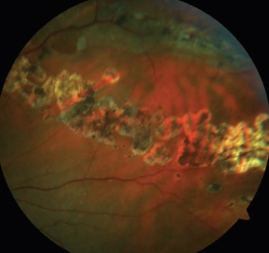
Lattice degeneration refers to thinning and weakening of the retina, often appearing as a lattice-like pattern of white lines or spots. This condition typically occurs in the peripheral retina, the outer edges of the retina, and can affect one or both eyes.

Symptoms:
- Floaters: Small, dark spots or lines that drift across the field of vision.
- Flashes of light: Brief flashes or flickers of light in the peripheral vision.
- Blurred vision: In severe cases, lattice degeneration can lead to distorted or blurred vision.
Risk Factors:
Several factors may increase the risk of developing lattice degeneration, including:
- Genetics: A family history of retinal disorders may predispose individuals to lattice degeneration.
- Nearsightedness, or myopia, increases the likelihood of lattice degeneration.
- Age: While lattice degeneration can occur at any age, it’s more common in individuals over 40.
Complications:
- Retinal tears: The weakened areas in the lattice pattern are more prone to tears, which can result in retinal detachment if left untreated.
- If a tear occurs and is not promptly treated, it can detach the retina from the back of the eye, causing severe vision loss or blindness.
Diagnosis:
Lattice degeneration is typically diagnosed during a comprehensive eye examination. Firstly, your eye doctor will dilate your pupils to obtain a clear view of the retina. Subsequently, they may use specialized imaging techniques, such as optical coherence tomography (OCT) or fundus photography, to assess the extent of the degeneration. In addition, these imaging methods help detect any associated complications, enabling accurate diagnosis and appropriate management.
Treatment:
- Laser therapy: Doctors can use laser photocoagulation to seal retinal tears and prevent further complications.
- In some cases, doctors may recommend cryotherapy, which involves freezing therapy, to treat retinal tears or strengthen the weakened areas of the retina.
- Cryotherapy: Doctors may recommend freezing therapy (cryotherapy) to treat retinal tears or strengthen weakened areas of the retina.
- Vitrectomy: In advanced cases of retinal detachment, a surgical procedure called vitrectomy may be necessary to reattach the retina.
Prevention:
- Regular eye exams: Routine eye examinations can help detect lattice degeneration and other retinal disorders early, allowing for prompt treatment.
- Wearing protective eyewear can help prevent retinal tears when engaging in activities that pose a risk of eye injury, such as contact sports or DIY projects.
- Manage underlying conditions: Keeping conditions like myopia under control through corrective lenses or refractive surgery may help reduce the risk of lattice degeneration progression.
Author Details:
Dr. Sushruth Appajigowda holds a prominent position as a cornea, cataract, glaucoma, and LASIK surgeon in Bangalore. He serves as the chief cataract and refractive surgeon at Vijaya Nethralaya Eye Hospital, Nagarbhavi, Bangalore. Renowned as one of the finest LASIK surgeons nationwide, he brings with him over 12 years of experience across multiple LASIK platforms, including ZEISS, ALCON, SCHWIND, AMO, and Bausch and Lomb. Having successfully conducted over 5000 LASIK procedures, Dr. Sushruth holds the title of a Certified Refractive Surgeon and a Fellow of the All India Collegium of Ophthalmology. Furthermore, he stands as a distinguished speaker at various national and international forums, using his expertise to guide you in selecting the most suitable procedure based on your health requirements.

https://vijayanethralaya.com/link-in-bio/
Conclusion:
While it may not always cause symptoms, it can increase the risk of retinal tears and detachment, leading to vision loss if left untreated. Regular eye exams and early intervention are crucial for managing lattice degeneration and minimizing the risk of complications.


