The Fundus test is one of the most important diagnostic procedures in modern ophthalmology. It plays a vital role in detecting retinal disorders, optic nerve abnormalities, vascular changes, and even systemic diseases such as diabetes and hypertension. In simple terms, this test allows doctors to look inside the back of your eye and assess its overall health.
In today’s world, where screen time is increasing and lifestyle-related diseases are becoming more common, regular eye examinations are more important than ever. A Fundus test helps identify issues early, often before symptoms appear. Early detection means early treatment—and that can make all the difference in preserving vision.
In this comprehensive guide, you will learn everything about the Fundus test, including its purpose, procedure, benefits, risks, interpretation of results, and frequently asked questions.
What Is a Fundus Test?
A fundus test, also known as a fundus examination or ophthalmoscopy, is a medical procedure that evaluates the interior surface of the eye. This includes:
- The retina
- The optic disc
- The macula
- Retinal blood vessels
- The posterior pole
The term “fundus” refers to the interior surface at the back of the eye. During the test, an eye specialist (ophthalmologist or optometrist) uses specialized equipment to visualize these structures.
This examination is painless, quick, and extremely informative. It helps doctors assess not only eye health but sometimes overall systemic health as well.

Why Is a Fundus Test Important?
It is essential because many eye diseases develop silently. Patients may not notice symptoms until significant damage has occurred. Early detection allows for timely treatment and prevention of vision loss.
Key Reasons for a Fundus Test
- Detection of Diabetic Retinopathy
Patients with diabetes are at high risk for retinal damage. Fundus examination identifies early changes in blood vessels. - Monitoring Hypertensive Retinopathy
High blood pressure affects retinal vessels. The test helps assess severity. - Glaucoma Evaluation
The optic nerve is examined for signs of glaucoma-related damage. - Macular Degeneration Screening
Age-related macular degeneration can be identified early. - Retinal Detachment Detection
Early signs of retinal tears or detachment can be spotted. - Assessment of Papilledema
Swelling of the optic disc may indicate increased intracranial pressure.
Clearly, the Fundus test is more than just a routine check—it is a powerful diagnostic tool.
Types of Fundus Examination
There are several methods used to perform a Fundus test. Each has specific advantages depending on the clinical situation.
1. Direct Ophthalmoscopy
This is a handheld device that provides a magnified view of the retina. It is commonly used in routine examinations.
Advantages:
- Quick and convenient
- Useful in primary care settings
Limitations:
- Limited field of view
2. Indirect Ophthalmoscopy
This technique uses a light source and a special lens to provide a wider field of view.
Advantages:
- Better visualization of peripheral retina
- Useful for detecting retinal tears
Limitations:
- Requires more training
3. Fundus Photography
Digital imaging captures detailed pictures of the retina. These images can be stored and compared over time.
Benefits:
- Excellent for documentation
- Useful for telemedicine
- Allows disease progression monitoring
4. Optical Coherence Tomography (OCT)
Although technically different, OCT is often associated with fundus evaluation. It provides cross-sectional images of retinal layers.
Best for:
- Macular diseases
- Diabetic macular edema
- Retinal thickness assessment
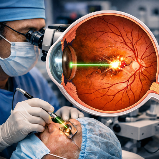
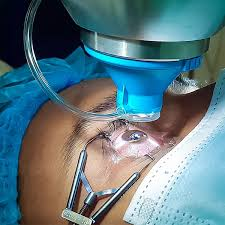
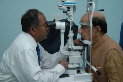
How Is a Fundus Test Performed?
It is simple and usually completed within 10–20 minutes.
Step-by-Step Procedure
- Patient Preparation
- The patient sits comfortably in a darkened room.
- Pupil-dilating eye drops may be administered.
- Pupil Dilation
- Dilating drops widen the pupil.
- This allows better visualization of the retina.
- Effects last 3–6 hours.
- Examination
- The doctor uses an ophthalmoscope or imaging device.
- The retina and optic nerve are examined carefully.
- Image Capture (if required)
- Digital photographs may be taken.
The procedure is painless. Some patients may feel mild discomfort from bright light exposure, but it is temporary.
Who Should Get a Fundus Test?
While everyone benefits from periodic eye exams, certain individuals require more frequent fundus evaluation.
High-Risk Groups
- Diabetic patients
- People with high blood pressure
- Individuals over 40 years old
- Patients with glaucoma risk
- Those with sudden vision changes
- People with family history of retinal disease
Global ophthalmology guidelines recommend annual screening for diabetic patients.
What Can a Fundus Test Detect?
It is capable of identifying numerous eye and systemic conditions.
Common Eye Conditions Detected
| Condition | What the Test Reveals |
|---|---|
| Diabetic Retinopathy | Microaneurysms, hemorrhages |
| Glaucoma | Optic nerve cupping |
| Macular Degeneration | Drusen deposits |
| Retinal Detachment | Tears or separation |
| Retinitis Pigmentosa | Pigment changes |
Systemic Conditions Identified
Interestingly, the retina is the only place where blood vessels can be directly visualized. That makes the fundus test valuable for detecting the following:
- Hypertension
- Atherosclerosis
- Stroke risk indicators
- Brain tumors (via papilledema)
Are There Any Risks or Side Effects?
It is extremely safe. However, mild temporary side effects may occur:
- Light sensitivity after dilation
- Blurred vision for a few hours
- Mild headache
- Rare allergic reaction to drops
Patients are usually advised not to drive immediately after dilation.
How to Prepare for a Fundus Test
Preparation is minimal, but a few steps can improve comfort:
- Bring sunglasses for post-exam light sensitivity
- Arrange transportation if dilation is planned
- Inform your doctor about medications
- Report any allergies
There is no need for fasting or special dietary restrictions.
Understanding Fundus Test Results
After the examination, the doctor explains the findings.
Normal Findings Include the following:
- Clear retina
- Sharp optic disc margins
- Healthy macula
- Normal blood vessel appearance
Abnormal Findings May Show:
- Swelling
- Hemorrhages
- Exudates
- Optic nerve damage
- Retinal thinning
Further testing may be recommended if abnormalities are detected.
Fundus Test in Diabetic Patients
Diabetes is one of the leading causes of blindness worldwide. According to the World Health Organization (WHO), diabetic retinopathy is a major global health concern.
Regular fundus test screening helps the following:
- Detect early retinal changes
- Prevent severe vision loss
- Monitor disease progression
- Guide treatment decisions
Laser therapy, injections, or surgery may be recommended based on findings.
Technology Advancements in Fundus Examination
Modern advancements have transformed the Fundus test into a highly precise diagnostic tool.
Recent innovations include the following:
- AI-assisted retinal screening
- Ultra-widefield imaging
- Portable fundus cameras
- Teleophthalmology integration
These technologies improve accessibility, especially in rural areas.
Conclusion
The Fundus test is a powerful and essential diagnostic tool that plays a central role in protecting vision and detecting systemic diseases. It is safe, quick, and highly informative. Regular fundus examinations can prevent serious eye complications, particularly in high-risk individuals such as diabetics and hypertensive patients.
Early detection truly saves sight. If you haven’t had an eye examination recently, scheduling a Fundus test could be one of the most important health decisions you make.
Taking proactive steps today ensures clearer vision tomorrow.
Frequently Asked Questions (FAQs)
1. Is a Fundus test painful?
No, the test is painless. You may feel temporary discomfort from bright light.
2. How long does pupil dilation last?
Dilation effects typically last 3 to 6 hours.
3. Can a Fundus test detect brain problems?
Yes, optic disc swelling may indicate increased intracranial pressure.
4. How often should I get a Fundus test?
Healthy adults should have regular eye exams every 1–2 years. Diabetic patients need annual screening.
5. Can children undergo a Fundus test?
Yes, especially if they have visual complaints or systemic conditions.