Introduction to Fundus Photography
What Is Fundus Photography?
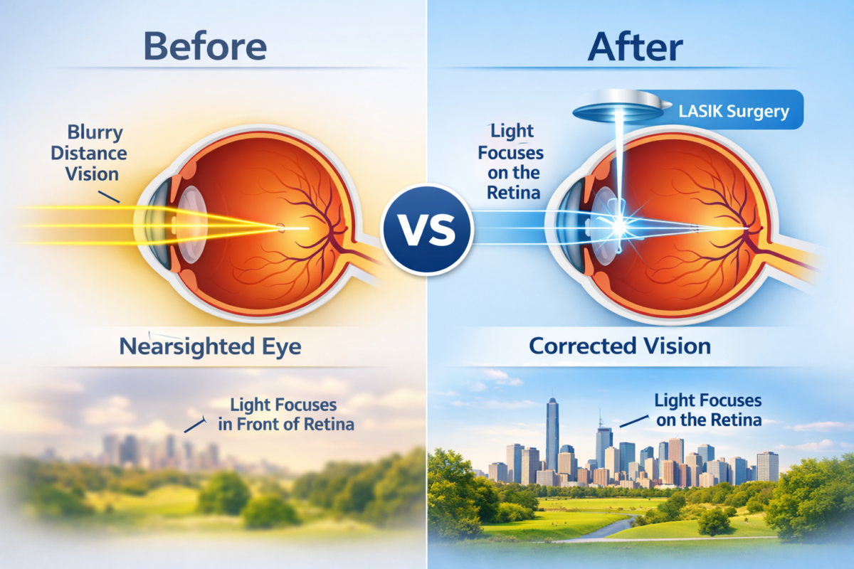
It is a specialized medical imaging technique used to capture detailed photographs of the interior surface of the eye. This area, known as the fundus, includes the retina, optic disc, macula, and blood vessels. These structures play a critical role in vision and overall eye health.
Think of fundus photography like taking a high-resolution landscape photo—except the landscape is inside your eye. With advanced cameras and lighting systems, eye specialists can visualize the retina in incredible detail.
This imaging method is widely used by ophthalmologists and optometrists to detect, document, and monitor various eye diseases. Because the retina reflects changes caused by systemic illnesses such as diabetes or hypertension, it often provides valuable insights into a person’s overall health.
In modern eye care, it has become a standard diagnostic tool. It allows doctors to compare images over time, making it easier to track disease progression and treatment effectiveness.
Why Retinal Imaging Matters in Modern Eye Care
Our eyes are incredibly complex organs, and many serious eye conditions develop silently without noticeable symptoms in their early stages. That’s where it becomes extremely valuable.
Here are some key reasons retinal imaging is essential:
- Early detection of eye diseases
- Monitoring chronic conditions
- Accurate medical documentation
- Remote diagnosis through telemedicine
Conditions such as Diabetic Retinopathy or Glaucoma can damage vision permanently if left untreated. Fundus photography helps doctors spot early warning signs before significant vision loss occurs.

History and Evolution of Fundus Photography
Early Retinal Imaging Techniques
The journey of fundus photography began in the mid-19th century when scientists attempted to capture images of the retina using rudimentary cameras. Early attempts were challenging due to poor lighting, long exposure times, and limited photographic technology.
The first successful retinal photograph was captured in 1886 by ophthalmologist Jackman and Webster. While the image quality was far from perfect, it marked the beginning of retinal imaging in medical science.
For decades afterward, ophthalmologists relied heavily on direct observation through ophthalmoscopes rather than photographic documentation.
Digital Transformation in Ophthalmology
The real breakthrough came with the development of digital fundus cameras in the late 20th century. Digital imaging revolutionized eye care by allowing:
- Instant image capture
- High-resolution retinal photos
- Easy storage and sharing of images
- Advanced software analysis
Today, digital fundus photography is used in hospitals, eye clinics, research institutions, and even mobile screening programs.
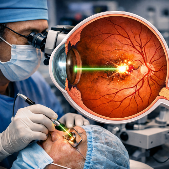
How Fundus Photography Works
Structure of the Human Retina
The retina is a thin layer of tissue at the back of the eye that converts light into electrical signals for the brain. When light enters the eye, it passes through the lens and focuses on the retina.
Important structures captured during fundus photography include:
- Optic disc – where the optic nerve exits the eye
- Macula – responsible for sharp central vision
- Retinal blood vessels
- Peripheral retina
These areas reveal important clues about eye health.
Key Parts Captured in Fundus Images
A typical fundus photograph shows:
| Structure | Function |
|---|---|
| Retina | Light detection |
| Macula | Central vision |
| Optic Disc | Connection to brain |
| Blood Vessels | Circulation |
Doctors analyze these features to detect abnormalities.
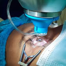
The Imaging Process Explained
It is the process is quick and painless. It usually involves these steps:
- The patient sits in front of the fundus camera.
- Sometimes eye drops are used to dilate the pupil.
- The patient focuses on a target light.
- The camera captures a flash image of the retina.
The entire procedure typically takes only a few minutes.
Types of Fundus Photography
Traditional Fundus Cameras
Traditional fundus cameras require pupil dilation to obtain clear images. These cameras provide highly detailed photographs and are commonly used in ophthalmology clinics.
They remain the gold standard for retinal imaging.
Non-Mydriatic Fundus Photography
Non-mydriatic fundus photography allows imaging without dilating the pupil. This technique is faster and more comfortable for patients.
Advantages include:
- Quick screening
- Minimal patient preparation
- Ideal for large screening programs
Ultra-Widefield Fundus Imaging
Ultra-widefield imaging captures up to 200 degrees of the retina in a single image. This provides a much broader view compared to standard fundus photography.
It is particularly useful for detecting peripheral retinal diseases.
Equipment Used in Fundus Photography
Fundus Camera Components
A typical fundus camera consists of several key parts:
- Optical system
- Illumination system
- Digital sensor
- Computer interface
These components work together to produce detailed retinal images.
Software and Image Analysis Tools
Modern fundus photography systems include advanced software that helps with:
- Image enhancement
- Disease detection
- Patient record management
- AI-based analysis
These tools help doctors make faster and more accurate diagnoses.
Medical Conditions Detected Using Fundus Photography
Diabetic Retinopathy
Diabetic Retinopathy is one of the leading causes of blindness worldwide. It occurs when high blood sugar damages retinal blood vessels.
It helps detect early signs such as:
- Microaneurysms
- Retinal hemorrhages
- Exudates
Early detection significantly reduces the risk of vision loss.
Glaucoma
Glaucoma damages the optic nerve, often due to increased eye pressure.
Fundus photographs allow doctors to examine the optic disc for changes such as cupping, which indicates nerve damage.
Age-Related Macular Degeneration
Age-related Macular Degeneration (AMD) affects the macula and causes central vision loss.
Fundus imaging helps identify:
- Drusen deposits
- Pigment changes
- Retinal damage
Hypertensive Retinopathy
High blood pressure can cause changes in retinal blood vessels. Fundus photography helps detect narrowing, leakage, or swelling in these vessels.
Role of Fundus Photography in Preventive Eye Care
Early Disease Detection
One of the biggest advantages of fundus photography is early detection. Many eye diseases progress silently.
Regular retinal imaging helps doctors detect problems before symptoms appear.
Monitoring Disease Progression
Doctors often compare fundus images taken over time to monitor disease progression. This helps determine whether treatments are working effectively.
Benefits of Fundus Photography
Non-Invasive Eye Examination
It is completely non-invasive. There are no needles, no surgery, and minimal discomfort.
Improved Diagnostic Accuracy
High-resolution images allow ophthalmologists to detect subtle retinal changes that might otherwise go unnoticed.
Patient Education and Documentation
Patients can actually see images of their own retina. This helps them better understand their condition and treatment plan.
Limitations and Challenges
Image Quality Issues
Poor image quality can occur due to:
- Small pupils
- Eye movement
- Cataracts
These factors can make interpretation difficult.
Cost and Accessibility
Advanced retinal imaging systems can be expensive. In some regions, access to fundus photography equipment remains limited.
Fundus Photography vs Other Retinal Imaging Techniques
Optical Coherence Tomography (OCT)
OCT provides cross-sectional images of the retina, allowing doctors to see its layers in detail.
While fundus photography shows surface images, OCT reveals internal retinal structure.
Fluorescein Angiography
This imaging technique uses a dye injected into the bloodstream to visualize retinal blood flow.
It is simpler and safer since it does not require injections.
Artificial Intelligence in Fundus Photography
Automated Disease Detection
Artificial intelligence is transforming fundus photography. AI algorithms can analyze retinal images and detect diseases with remarkable accuracy.
For example, AI systems can screen thousands of images for diabetic retinopathy in seconds.
Future AI-Driven Diagnostics
AI will likely play an even bigger role in ophthalmology by:
- Predicting disease risk
- Assisting doctors with diagnosis
- Enabling remote eye screening
Organizations such as the World Health Organization highlight digital screening as a key strategy for preventing blindness globally.
Future Trends in Fundus Photography
Tele-ophthalmology
Telemedicine allows retinal images to be sent to specialists remotely. This makes eye care accessible in rural or underserved regions.
Smartphone-Based Fundus Cameras
Portable smartphone fundus cameras are emerging as affordable alternatives to traditional systems.
These devices may revolutionize eye screening worldwide.
Conclusion
It has become one of the most powerful tools in modern ophthalmology. By capturing detailed images of the retina, this technology allows doctors to detect eye diseases early, monitor treatment progress, and improve patient outcomes.
From traditional fundus cameras to AI-powered diagnostic systems, retinal imaging continues to evolve rapidly. As technology advances, it will play an even greater role in preventive eye care and global blindness prevention.
Simply put, a small photograph of the retina can reveal a world of information about your eye health—and sometimes even your overall health.
Author Details:
Dr. Pavan K P is a highly experienced Ophthalmologist/Eye Surgeon, with over 20 years of experience under his belt. After he completed his MBBS from JSS Medical College in 1997, he went on to do his MS in Ophthalmology from Annamalai University in 2003, and later pursued his Fellowship in Vitreoretinal Surgery from Rajiv Gandhi University in 2005. Dr. Pavan K P is an expert when it comes to ROP Screening, Retinal Detachment Surgery, Laser Procedures for Retinal Diseases, Retinal Diseases, Diabetic Eye checkups, and Avastin Injections.
And he practices at Vijaya Nethralaya in Nagarbhavi, Bangalore. You can book an online appointment to consult with him at www.vijayanethralaya.com. Find his contact information and write a review of your experience on the website.

Dr. Pavan K P
Book your appointment
Frequently Asked Questions About Fundus Photography
1. Is fundus photography painful?
No. Fundus photography is painless and non-invasive. Patients may only experience a brief flash of light.
2. How long does a fundus photography test take?
The procedure usually takes 5–10 minutes, depending on whether pupil dilation is required.
3. Do I need to dilate my eyes for fundus photography?
Not always. Some cameras use non-mydriatic technology that works without dilation.
4. Can fundus photography detect systemic diseases?
Yes. Conditions like diabetes and hypertension often show signs in retinal blood vessels.
5. How often should fundus photography be done?
Patients with diabetes or other eye conditions may need annual retinal imaging. Your eye doctor will recommend the appropriate schedule.
6. Is fundus photography safe?
Yes. It uses safe light flashes and poses no health risks.