What Is a Fundus Eye Test?
A fundus eye test is a medical eye examination that allows doctors to examine the inside surface of your eye, known as the fundus. This area includes the retina, optic disc, macula, and retinal blood vessels. By looking at these structures, eye specialists can detect early signs of many serious eye and health conditions.
Think of the test as a “window into your health.” The blood vessels and nerves inside your eye can reveal problems not only related to vision but also systemic diseases like diabetes or high blood pressure.
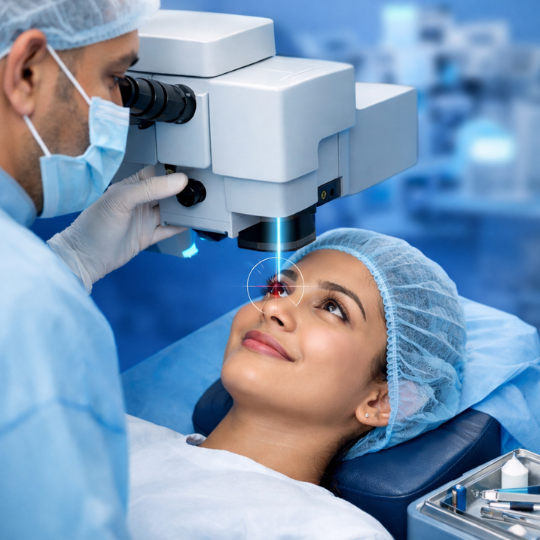
The test is usually performed by an ophthalmologist or optometrist using specialized equipment that magnifies and illuminates the back of the eye.
Understanding the Structure of the Eye Fundus
The fundus consists of several critical parts:
- Retina —a light-sensitive layer that converts light into signals sent to the brain.
- Macula —responsible for sharp central vision.
- Optic nerve – Transfers visual information to the brain.
- Blood vessels supply oxygen and nutrients to eye tissues.
These structures work together to produce clear vision. Any damage or abnormality here can lead to serious visual problems.
How the Test Helps Detect Eye Diseases
The biggest advantage of the Fundus Eye Test is early detection. Many eye diseases develop silently without noticeable symptoms.
For example, conditions like diabetic retinopathy or glaucoma can progress for years before vision problems appear. With a fundus examination, doctors can identify early changes and begin treatment immediately.
Why Doctors Recommend a Fundus Eye Test
Doctors often recommend this test for both preventive care and diagnosis, as it can help detect conditions such as glaucoma, diabetic retinopathy, and macular degeneration early on.
Routine Eye Health Screening
Even if you have perfect vision, routine eye examinations are important. A fundus check helps doctors confirm that the retina and optic nerve are healthy.
Early detection prevents permanent vision damage.
Monitoring Chronic Conditions
People with certain health conditions require regular retinal monitoring.
Common examples include:
- Diabetes
- High blood pressure
- Cardiovascular diseases
- Neurological disorders
Damage to retinal blood vessels may indicate complications from these diseases.
Conditions Detected Through a Fundus Eye Test
One reason doctors rely on this test is its ability to reveal multiple eye diseases in a single exam.
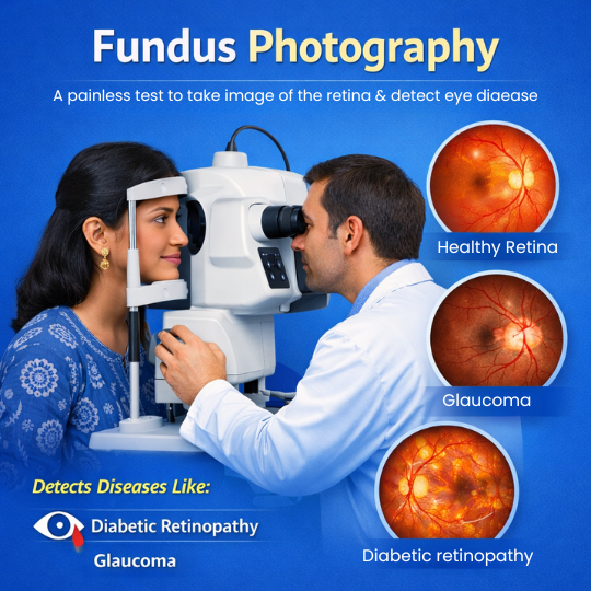
Diabetic Retinopathy
Diabetic retinopathy occurs when high blood sugar damages retinal blood vessels. Small leaks, swelling, or bleeding can appear in the retina.
Without treatment, it may lead to blindness.
Fundus imaging allows doctors to detect:
- Microaneurysms
- Retinal hemorrhages
- Abnormal blood vessel growth
Glaucoma
Glaucoma is a condition that damages the optic nerve due to high eye pressure.
During a fundus exam, doctors check the optic disc for:
- Enlargement
- Changes in color
- Structural damage
These changes help diagnose glaucoma early.
Macular Degeneration
Age-related macular degeneration, a condition that affects the central vision needed for reading and recognizing faces, is a common eye disorder in older adults.
The test reveals:
- Yellow deposits called drusen
- Retinal thinning
- Abnormal blood vessels
Retinal Detachment
Retinal detachment is a serious emergency where the retina separates from the underlying tissue.
Symptoms may include:
- Sudden flashes
- Floaters
- Shadow in vision
A fundus exam allows doctors to quickly identify tears or detachment.
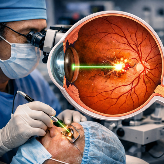
Types of Fundus Eye Tests
Several techniques are used depending on the patient’s condition and diagnostic needs.
Direct Ophthalmoscopy
This is the simplest method. A handheld device called an ophthalmoscope shines light into the eye.
Advantages:
- Quick
- Non-invasive
- Useful for routine checks
However, it provides a limited field of view.
Indirect Ophthalmoscopy
Indirect ophthalmoscopy uses a head-mounted light and a special lens.
Benefits include:
- Wider retinal view
- Better depth perception
- Detection of peripheral retinal problems
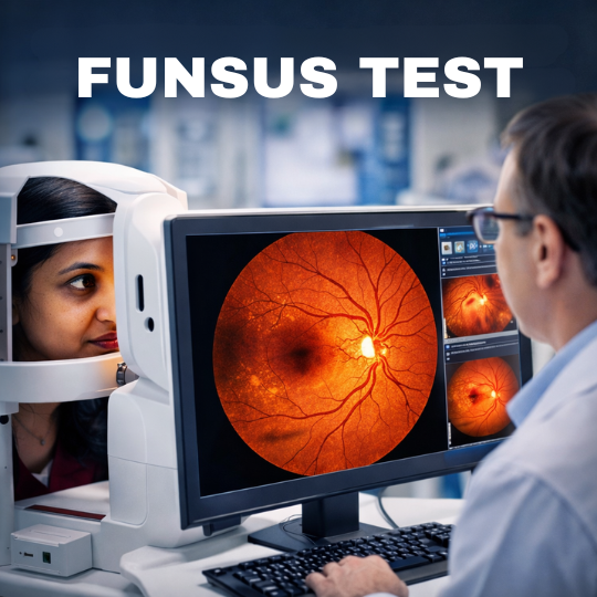
Fundus Photography
Fundus Photography captures detailed digital images of the retina.
These images help doctors:
- Track disease progression
- Compare previous results
- Share reports with specialists
Many modern clinics use high-resolution retinal cameras for this purpose.
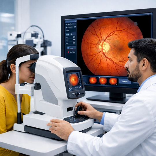
Step-by-Step Procedure of a Fundus Eye Test
Many patients feel nervous before medical tests, but this exam is simple and painless.
Preparation Before the Test
Before the examination:
- Eye drops may be used to dilate the pupils
- This allows the doctor to see deeper into the retina
- Vision may become temporarily blurry
It’s recommended to bring sunglasses because light sensitivity may occur.
During the Examination
During the test:
- You sit in front of the examination device.
- The doctor shines a light into your eye.
- They carefully inspect the retina and optic nerve.
- Images may be captured using retinal cameras.
The procedure usually takes 5–15 minutes.
After the Test
Once the exam is complete:
- Your pupils may stay dilated for several hours
- Reading or driving might be difficult temporarily
- Vision returns to normal after the drops wear off
Doctors may discuss the results immediately or schedule a follow-up if abnormalities appear.
Benefits of Getting a Fundus Eye Test
Regular retinal examinations provide many health advantages.
Key benefits include the following:
- Early detection of vision-threatening diseases
- Monitoring systemic diseases like diabetes
- Preventing irreversible blindness
- Tracking treatment progress
- Detecting neurological issues
According to the World Health Organization, early eye disease detection significantly reduces the risk of preventable blindness.
Who Should Get a Fundus Eye Test?
While everyone can benefit from the exam, some people should prioritize it.
High-risk groups include:
- Adults over 40
- People with diabetes
- Individuals with high blood pressure
- Those with a family history of eye disease
- People experiencing sudden vision changes
Children may also undergo the test if doctors suspect retinal abnormalities.
Risks or Side Effects
The procedure is generally very safe.
Possible temporary effects include:
- Light sensitivity
- Blurred vision from dilating drops
- Mild eye discomfort
These symptoms usually disappear within a few hours.
Serious complications are extremely rare.
Fundus Eye Test vs Regular Eye Exam
Many people assume both tests are the same, but they serve different purposes.
| Feature | Regular Eye Exam | Fundus Eye Test |
|---|---|---|
| Purpose | Vision correction | Retinal health |
| Focus | Glasses prescription | Internal eye structures |
| Tools | Vision charts | Ophthalmoscope or retinal camera |
| Diseases detected | Refractive errors | Retinal diseases |
Both tests are essential for complete eye care.
How Often Should You Take a Fundus Eye Test?
The recommended frequency depends on age and health.
General guidelines:
- Healthy adults: every 1–2 years
- Diabetic patients: annually
- Older people over 60: yearly
- People with eye disease: as recommended by a doctor
Regular screening dramatically improves treatment success.
Tips to Maintain Healthy Eyes
Eye care goes beyond medical exams.
Here are some practical tips:
- Eat foods rich in vitamin A and omega-3s.
- Take breaks from screens (20-20-20 rule)
- Wear UV-protective sunglasses
- Avoid smoking
- Maintain healthy blood sugar levels
Healthy lifestyle choices protect your retina and vision.
Future of Eye Diagnostics
Eye care technology is advancing rapidly.
Emerging innovations include:
- AI-assisted retinal screening
- Smartphone retinal imaging
- Tele-ophthalmology for remote diagnosis
- Advanced 3D retinal scans
These tools make early detection easier and more accessible worldwide.
Conclusion
Vision is one of our most valuable senses, yet many people ignore eye health until problems appear. A fundus eye test is a simple but powerful examination that allows doctors to detect serious eye diseases long before symptoms develop.
By examining the retina, optic nerve, and blood vessels, specialists can diagnose conditions such as diabetic retinopathy, glaucoma, and macular degeneration early. The test is quick, painless, and incredibly valuable for protecting long-term vision.
Regular eye checkups combined with healthy lifestyle habits can significantly reduce the risk of vision loss. If you haven’t had a retinal exam recently, scheduling one could be one of the smartest decisions you make for your eye health.
Author Details:
Dr. Pavan K P is a highly experienced Ophthalmologist/Eye Surgeon, with over 20 years of experience under his belt. After he completed his MBBS from JSS Medical College in 1997, he went on to do his MS in Ophthalmology from Annamalai University in 2003, and later pursued his Fellowship in Vitreoretinal Surgery from Rajiv Gandhi University in 2005. Dr. Pavan K P is an expert when it comes to ROP Screening, Retinal Detachment Surgery, Laser Procedures for Retinal Diseases, Retinal Diseases, Diabetic Eye checkups, and Avastin Injections.
And he practices at Vijaya Nethralaya in Nagarbhavi, Bangalore. You can book an online appointment to consult with him at www.vijayanethralaya.com. Find his contact information and write a review of your experience on the website.

Dr. Pavan K P
Book your appointment
FAQs About Fundus Eye Test
1. Is a fundus eye test painful?
No. The test is completely painless. You may feel slight discomfort from bright light or dilating drops, but it lasts only a few minutes.
2. How long does the test take?
Most examinations take 5 to 15 minutes, depending on whether imaging is required.
3. Can the test detect brain problems?
Yes. Changes in the optic nerve or retinal blood vessels can sometimes indicate neurological conditions or increased brain pressure.
4. Do I need to prepare for the test?
Usually no preparation is required. However, bring sunglasses because dilating drops may cause temporary light sensitivity.
5. Can children undergo a fundus eye test?
Yes. Pediatric ophthalmologists perform the exam to detect congenital retinal issues or developmental eye disorders.
6. Is the test safe during pregnancy?
Yes. The examination itself is safe, though doctors may avoid certain medications unless necessary.