Introduction:
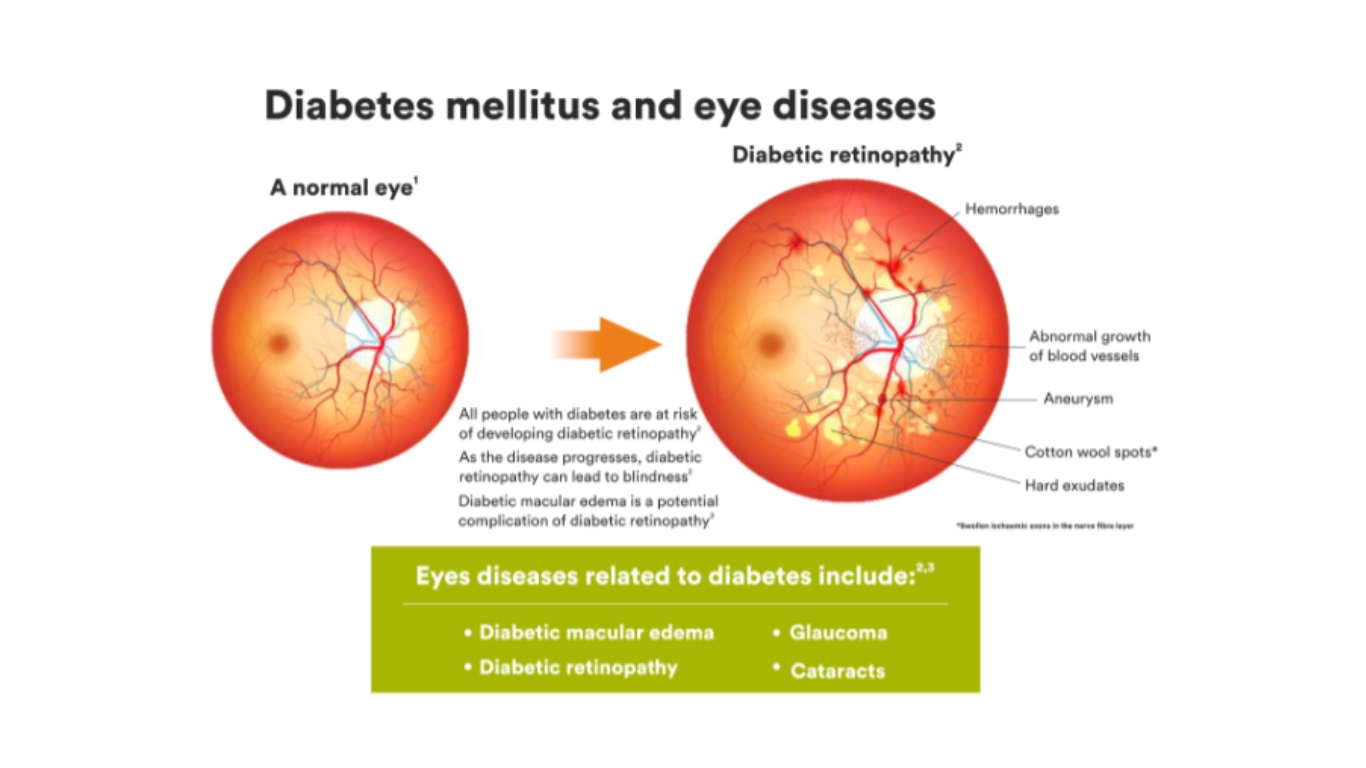
Diabetic macular edema (DME) is a condition that affects the eyes of individuals with diabetes. It occurs when fluid leaks into the macula, which is the central part of the retina responsible for sharp, central vision. As a result, the macula swells, leading to blurred or distorted vision. Moreover, DME commonly arises as a complication of diabetic retinopathy, a condition that affects blood vessels in the retina. Due to high blood sugar levels in diabetes, the blood vessels can sustain damage, causing leakage and the formation of abnormal blood vessels. Consequently, the fluid leakage into the macula is triggered, giving rise to the development of DME.
What is Diabetic Macular Edema (DME)?
Uncontrolled Diabetes Mellitus (DM) manifests as the accumulation of fluid in the central most part of the retina known as the macula. Retina is the light sensitive tissue at the back part of the eye and macula is the central part responsible for fine vision.
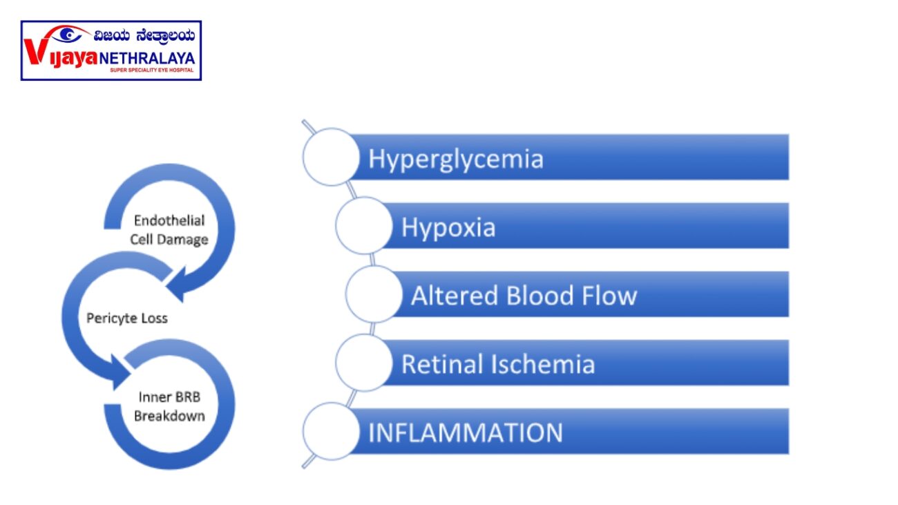
Pathophysiology-

What are the symptoms of DME?
- Blurring of central vision-Patients complains of blurry vision making it difficult to read and write, sew and do near work.
- Distorted central vision- this reduces the quality of vision, making it difficult to recognise faces and objects.
- The advanced stages of the condition typically exhibit a central black spot (scotoma). In the majority of cases, individuals typically observe the preservation of peripheral vision.

The diagnosis of DME (Diabetic Macular Edema):
The retina specialist can often detect it during a clinical examination.
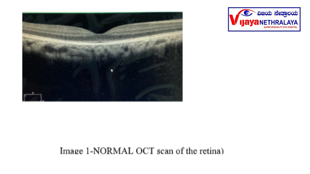
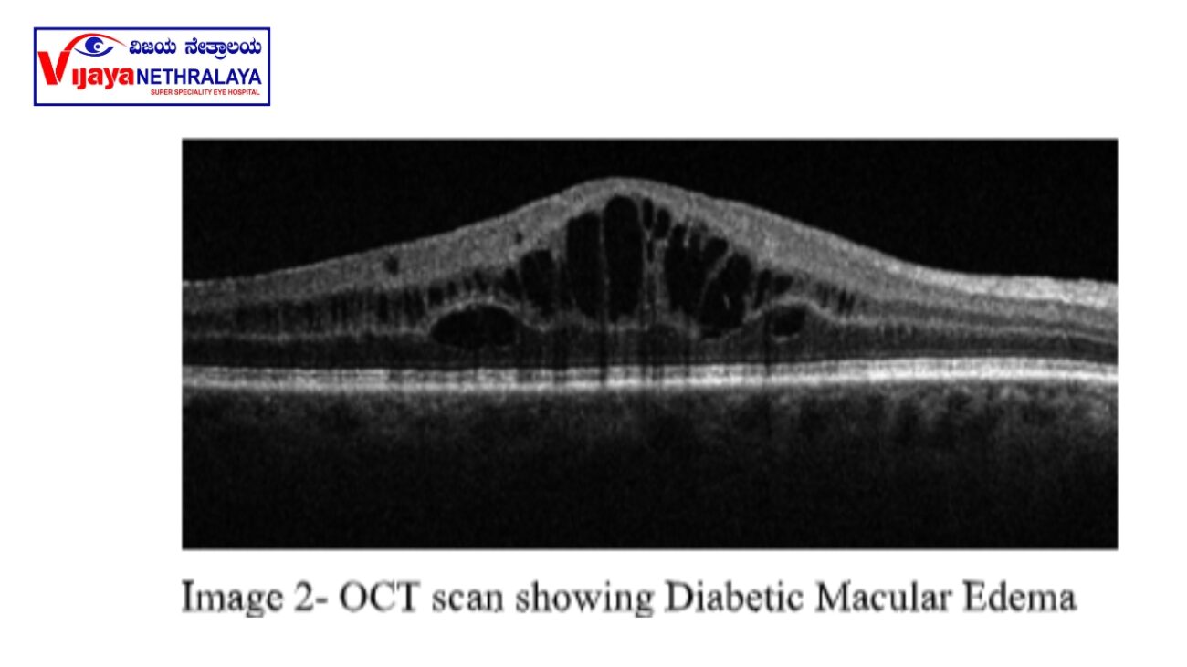
The healthcare provider recommends an Optical Coherence Tomography (OCT) scan of the retina as a means to quantify the amount of fluid, which proves useful when initiating treatment. Additionally, OCT captures high-resolution cross-sectional images (refer to images 1 and 2) of the central part of the retina known as the macula, where CNVMs are commonly observed. This quick and non-invasive test effectively images the retina. Furthermore, after the patient’s pupils are dilated, they are instructed to sit upright on a chair in front of the OCT machine, rest their chin on a chinrest, and undergo the scanning process (refer to image 3).
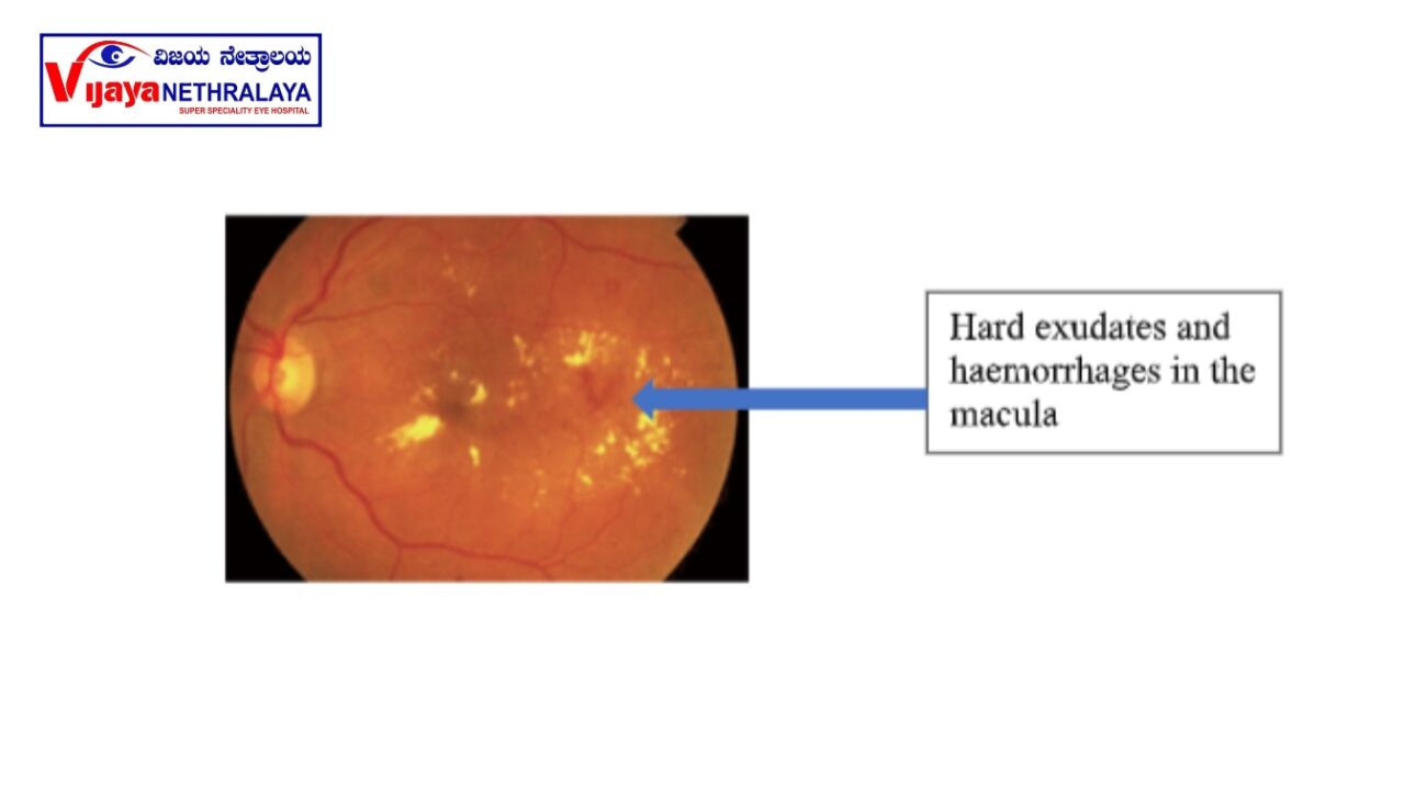
FUNDUS PHOTOS:
These are captured using a retinal camera to document the lesion and serve as a reference for comparison during follow-up examinations.
What is the treatment of DME?
ANTI VEGF INJECTIONS:
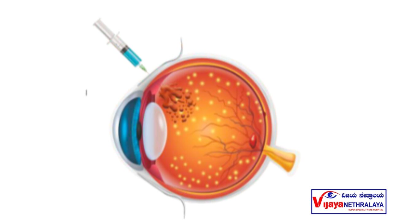
These are the main modality of treatment. It prevents vision loss, stabilizes vision, and improves vision if administered at the right time. These include drugs like Bevacizumab,
Ranibizumab, Aflibercept and Brolucizumab. They act by limiting the leakage from pathologic blood vessels, and control disease activity. The injections are administered as monthly or bimonthly doses, depending on the molecule and severity of the disease. Regular follow-up is necessary as advised by the treating retina specialist.
INTRAVITREAL STEROIDS:
In certain case scenarios, the retina specialist may advise steroid injections in the form of dexamethasone implants based on the appearance of DME. The advantage of these
implants is that it stays in the eye for a longer period resulting in less frequent injections. Some patients may develop high intraocular pressures and progression of cataracts post
injection, hence proper case selection and follow up is highly important.
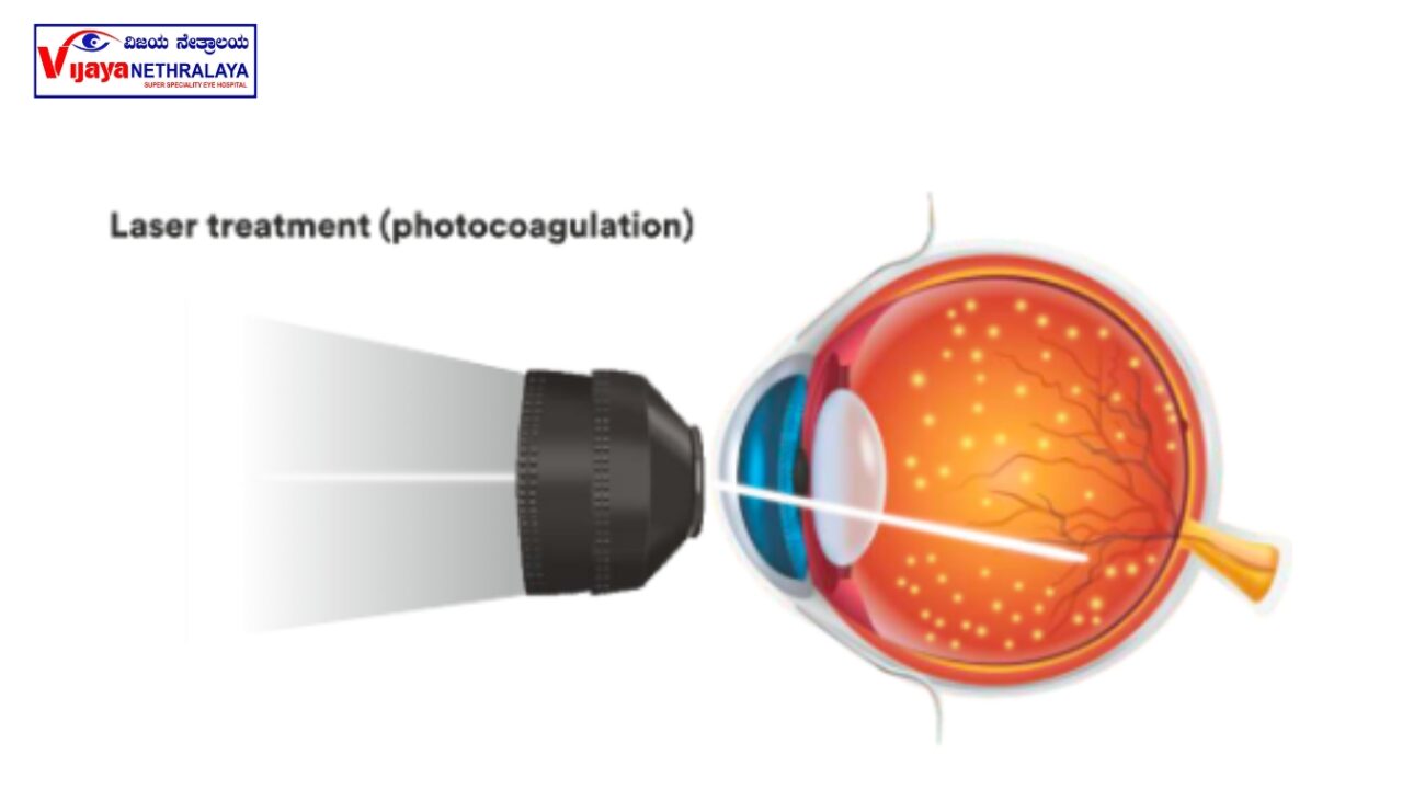
MACULAR LASER:
In some cases, the retina specialist will advise a macular laser. This procedure seals the leaking blood vessels and reduces DME. The procedure, conducted in an OPD setting, takes approximately 10-15 minutes to complete after dilation.
How can we prevent DME?
Strict control of Diabetes is the mainstay to prevent Diabetic complications in the retina which include DME. Chronic conditions such as high blood pressure, obesity or overweight, and high cholesterol can indirectly impact diabetic macular edema and its related complications.Maintain a healthy diet and regular exercise goes a long way in controlling DM. Avoid smoking. It is recommended to have regular retina check-ups, at least once a year, and to follow the advised schedule.
Author Details:
Dr. Sherina Thomas Expertise in diagnosing and treating all types of medical retinal diseases including Diabetic Retinopathy, Age-related macular degeneration, other vascular diseases of the retina with special interest in inherited retinal disorders.Surgically trained to manage Rhegmatogenous Retinal Detachment, Tractional Retinal Detachment, Macular Hole, Epiretinal Membrane, Severally fixated Intra ocular lens implantation.Expert in diagnostic modalities like Fundus Fluorescein Angiography, Indocyanine Angiography, Optical Coherence Tomography, B scan.

Conclusion:
In conclusion, diabetic macular edema (DME) is a common complication of diabetes that affects the macula, leading to blurred or distorted central vision. It occurs due to leakage of fluid from damaged blood vessels in the retina. Regular eye examinations are vital for early detection and monitoring of DME in individuals with diabetes.

