Introduction:
At Vijaya Nethralaya, we are proud to present the Aurolab Aqueous Drainage Implant, a revolutionary breakthrough in glaucoma treatment. In this article, we will discuss the features, benefits, and efficacy of our innovative implant, highlighting its superiority over existing solutions in the market. If you are searching for an effective and reliable treatment option for glaucoma, look no further than the Aurolab Aqueous Drainage Implant.
Understanding AUROLABAQUOUS DRAINAGE IMPLANT:
During the surgical procedure, ophthalmologists perform the Aurolab Aqueous Drainage Implant (AADI) on patients who have elevated eye pressure that does not respond to maximum medical therapy. The procedure involves placing an implant on the sclera, which is the white part of the eye. This implant incorporates a tube that the surgeon inserts inside the eye, allowing for the drainage of fluid from the inside to the outside of the eye. Within approximately 5-8 weeks, a capsule forms around the implant, facilitating controlled fluid drainage and preventing complications associated with low eye pressure.
Who are the right candidates for this surgery?
- Patients who are not responding maximal medical therapy
- Failed trabeculectomy
- Neovascular glaucoma
- Congenital glaucoma
- Uveitic glaucoma
- Other secondary glaucoma
Design of the Aurolab Aqueous Drainage Implant:
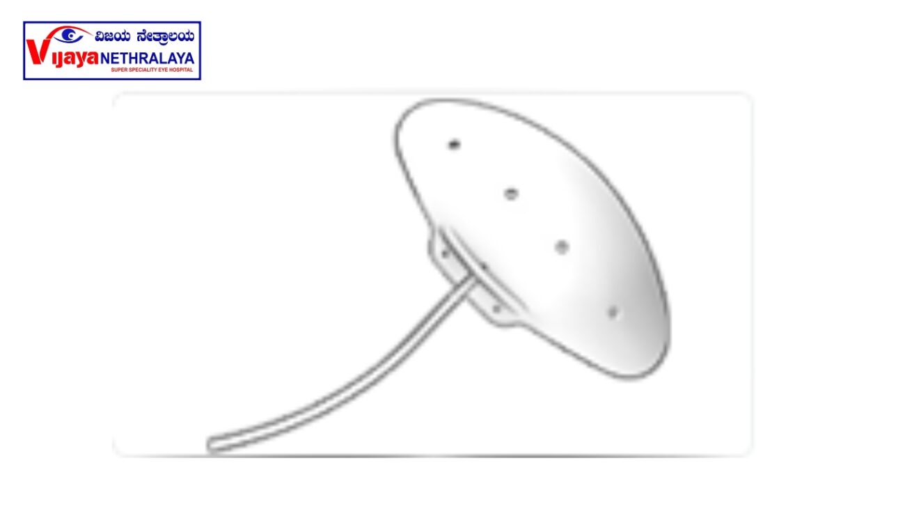
- Non-valved device.
- Made up of silicone material.
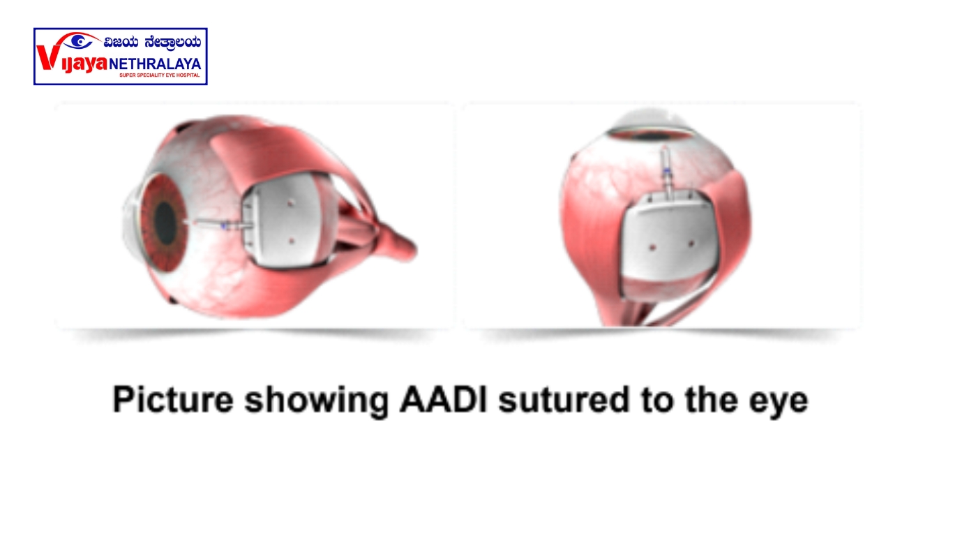
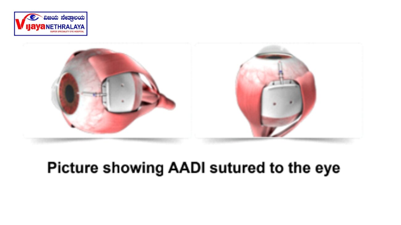
- The Aurolab Aqueous Drainage Implant (AADI) features a silicone plate with two fixation holes. These holes serve the purpose of securely suturing the plate to the sclera using 8-0 or 9-0 nylon sutures. This method of fixation ensures the stable positioning of the implant, allowing for effective fluid drainage and pressure management within the eye.
- It has a large surface area of 350 mm; this feature helps in effective filtration of aqueous fluid.
- When performing the surgical placement of the Aurolab Aqueous Drainage Implant (AADI), the surgeon strategically positions the device within a specific quadrant of the eye.
- Surgeons meticulously insert the lateral wings of the implant beneath the muscle in both the superior and temporal quadrants.
- This precise placement ensures optimal stability and effectiveness of the implant in managing intraocular pressure and promoting proper fluid drainage. This placement ensures both stability and cosmetic acceptability, as the device remains discreetly covered by the conjunctiva and eyelid.
- The AADI’s construction utilizing silicone material provides the surgeon with a high level of flexibility when handling the device. This flexibility enables precise maneuvering and placement, contributing to the overall success of the procedure.
- It has a unique fenestration design that allows the growth of fibrous tissue to control height and volume of the bleb and also to secure the device in place and minimize movements.
- This is much more affordable to the patients. AADI device.
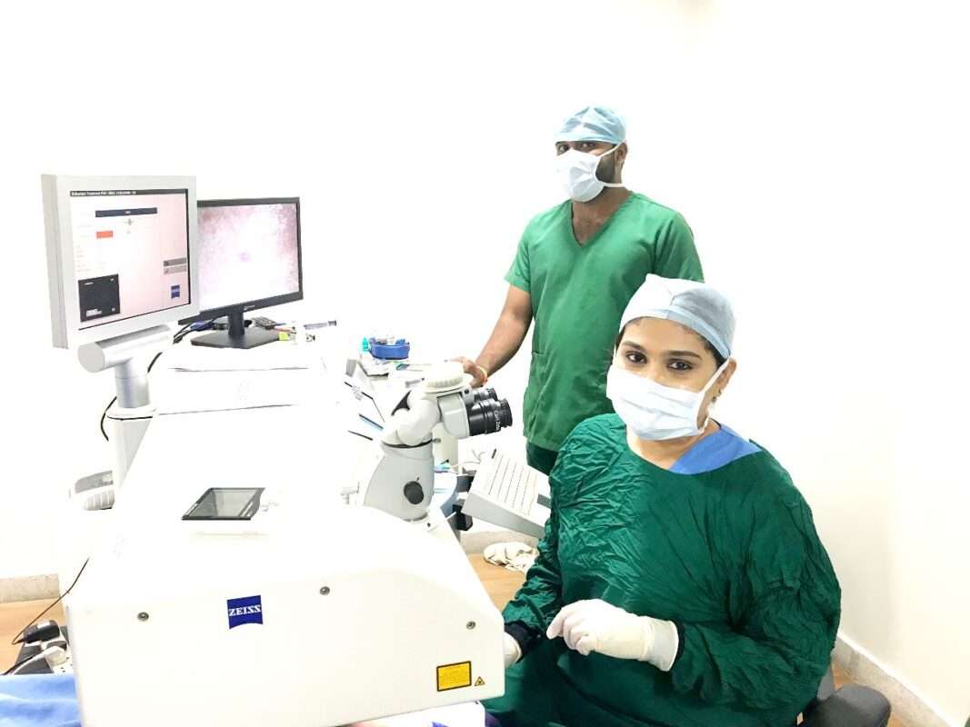
Performance of the Surgery:
- Your surgeon gives a local anesthesia to make your eye numbed during surgery and also to prevent any eye movements during surgery.
- The healthcare team positions you on the operating table and instructs you to lie down.
- The healthcare professional cleans and drapes your eye and then places a speculum to keep your eye open.
- By applying a stay suture on the cornea, the healthcare professional fixes the eyeball to one side.
- The healthcare professional cuts open the conjunctiva, which is the white part of the eye, and then fixes the device over the sclera and sutures to it.
- Before fixing the implant, the tube is always primed with sterile water and checked for patency.
- A small scleral tunnel is created through which the tube is inserted and entered into the eye.
- The tube is placed in such a way that it is short and not in contact with the cornea to prevent any damage to the surrounding structures.
- Your surgeon may or may not use a scleral patch graft to cover over the tube.
- With the help of sutures, the healthcare professional secures the tube to the underlying sclera.
- The healthcare professional covers the entire device and tube with the conjunctiva and sutures them in place.
Postoperative recovery:
- Your doctor puts you on antibiotic and steroid eye drops for 6–8 weeks.
- It is important for you to continue your anti-glaucoma medications for at least 5-6 weeks. This is the time when ligature resolves and capsule forms. After which, your surgeon tapers and stops the glaucoma eyedrops.
- You are expected to schedule postoperative eye pressure check-ups as advised by your surgeon.
What are the complications with this surgery?
- High or low eye pressure
- Bleeding inside the eye
- Eye infection
- Corneal edema
- Tube retraction
- Implant migration
How is AADI different from AGV surgery?
Both the procedures are glaucoma drainage devices, but they are different in the mechanism by which they drain the fluid. AADI is a non-valvular device, so your surgeon must ligate the tube with absorbable sutures, which disintegrate at around 5-6 weeks. This is the time when the fluid actually starts draining out, so that the capsule is already formed around the device. whereas AGV is a valve device, and this valve opens when the IOP is above 12 mmHG and closes when the IOP is around 8 mmHG. Here, the fluid is drained immediately after the surgery, and there is no need to ligate the tube like in AADI surgery.
Author Details:
Dr. Thanemozhi Srinivasan is an esteemed ophthalmologist who has achieved remarkable success in the specialized field of glaucoma. With a diverse and illustrious background, she has garnered extensive experience and honed her expertise in diagnosing, managing, and treating this intricate eye condition. Driven by a profound dedication to her patients, Dr. Srinivasan embraces cutting-edge techniques and innovative treatments, aiming to optimize visual outcomes while delivering compassionate and individualized care. With her wealth of knowledge, remarkable proficiency, and unwavering commitment to excellence, Dr. Thanemozhi Srinivasan stands as a revered authority in the realm of glaucoma.

Conclusion:
AADI is an implantable glaucoma drainage surgery done for any type of complicated glaucoma that is cost-effective for the patients. This is the best option since studies have shown a long-term success rate. This surgery is done only to control the eye pressure and not to reverse the damage that has already happened to the optic nerve.