Ocular Coherence Tomography (OCT) is a non-invasive, high-definition imaging test that uses light waves to capture live, cross-sectional pictures of your retina. Often described as an “optical ultrasound,” the OCT scan allows your eye doctor to see beneath the surface of the eye to map and measure the thickness of each individual retinal layer with microscopic precision.
Why This Guide Matters for Your Vision
If you have been scheduled for an OCT eye scan, you are about to undergo one of the most advanced diagnostic tests available in modern medicine. In the past, eye doctors could only see the surface of the retina. Today, Ocular Coherence Tomography (OCT) acts like a high-powered microscope that can “see” through the tissue, identifying microscopic changes long before you notice a change in your vision.
This guide is designed to walk you through every step of the process, explain the technology in simple terms, and help you understand why this test is the “gold standard” for preventing blindness.
How Does Ocular Coherence TomographyOCT Work? (The Science Made Simple)
To understand how OCT works, think of how a sonar or a traditional ultrasound works. An ultrasound sends sound waves into the body; those waves bounce off organs and return to a sensor to create a map.
OCT does the exact same thing, but it uses light waves instead of sound.
The “Bread Slice” Analogy
Imagine your retina is like a loaf of bread. A standard eye exam (where the doctor looks into your eye with a bright light) is like looking at the crust of the bread. You can see if the crust is burnt or cracked, but you don’t know what’s inside.
An OCT scan is like taking a slice out of that loaf. It allows the doctor to see the “texture” inside—the layers of nerves, blood vessels, and photoreceptors—without ever touching or cutting the eye.
Ocular Coherence Tomography (OCT)
What is an Ocular Coherence Tomography (OCT) Eye Scan Like? (The Patient Experience)
One of the biggest concerns patients have is whether the test will hurt or be uncomfortable. The answer is a definitive no.
The Procedure Step-by-Step
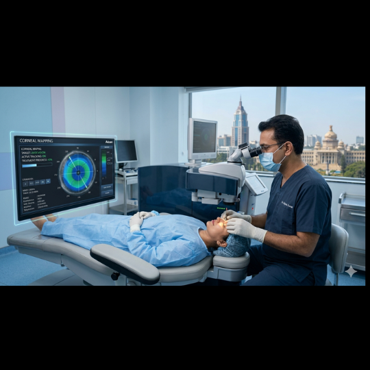
Check-in and Setup: You will be led to a darkened room where the OCT machine (which looks a bit like a large computer monitor with a chin rest) is located.
Positioning: You place your chin on the padded rest and your forehead against a bar. This keeps your eye perfectly still for the high-speed camera.
The “Target”: You will be asked to look at a small, green blinking light or a crosshair inside the machine.
The Capture: As you stare at the light, the machine’s “arm” will move close to your eye (but never touch it). You will see a flash of light or a red line sweep across your field of vision.
Duration: The actual scanning takes about 20 to 30 seconds per eye. The entire appointment usually lasts 10 minutes.
Will my eyes be dilated?
In many modern clinics, dilation is not strictly necessary for an OCT scan because the cameras are highly sensitive. However, if you have very small pupils or dense cataracts, your doctor may use “dilating drops” to enlarge your pupil, allowing more light into the eye for a clearer image.
What Happens During an Ocular Coherence Tomography (OCT Eye Scan?
One of the most common questions patients ask is, “What is an OCT eye scan like?” The good news is that the procedure is fast, painless, and requires no physical contact with your eye.
The Step-by-Step Process:
Preparation: You will sit in front of the OCT machine and rest your chin on a support to keep your head still.
Focus: You will be asked to look at a target light inside the machine.
The Scan: The machine will flash a light as it scans your eye. You won’t feel a thing.
Completion: The entire process usually takes about 5 to 10 minutes for both eyes.
Does it require dilation?
In many cases, your ophthalmologist can perform an OCT scan without dilating your pupils. However, if your pupils are very small or if the doctor needs a clearer view of the peripheral retina, they may use dilating drops.
Conditions Detected by OCT Imaging
An OCT scan is not just a routine check; it is a diagnostic powerhouse. It is specifically used to monitor the health of the macula (the center of your vision) and the optic nerve.
Condition
How OCT Helps
Glaucoma
Detects thinning of the optic nerve fibers before you lose sight.
Macular Degeneration (AMD)
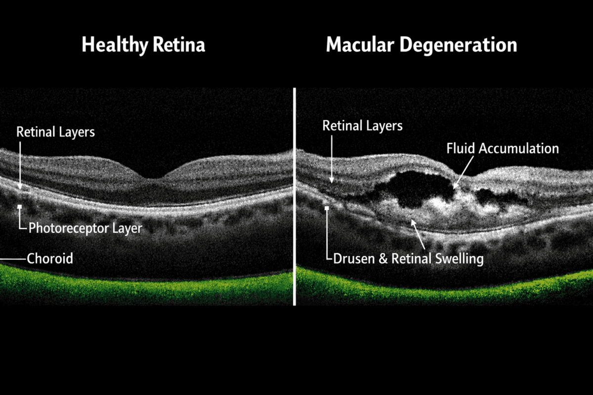
Identifies “drusen” (deposits) or fluid leakage under the retina.
Diabetic Retinopathy
Shows swelling in the retina (edema) caused by leaking blood vessels.
Macular Pucker/Hole
Visualizes physical pulling or tears in the retinal tissue.
Central Serous Retinopathy
Detects fluid buildup between retinal layers.
OCT for Glaucoma Detection
Glaucoma is a disease of the optic nerve. By the time you notice a “blind spot” from glaucoma, you have already lost 40% of your nerve fibers. OCT for glaucoma detection is a lifesaver because it measures the thickness of the nerve fiber layer. If the machine detects even a 1% thinning, your doctor can start treatment immediately.
Dry AMD: The OCT shows “drusen” (yellow deposits) lifting the retinal layers.
Wet AMD: The OCT shows actual fluid or blood leaking between the layers, appearing as dark “bubbles” on the scan.
Diabetic Retinopathy OCT Screening
Diabetes damages the small blood vessels in the eye. A diabetic retinopathy OCT screening detects “Macular Edema”—swelling caused by leaking fluid. Seeing this swelling on a scan allows for laser treatments or injections that can save your sight.
OCT retinal scan cross-section showing macular layers and fluid buildup in macular degeneration
OCT vs. Retinal Exam: Is One Better?
Patients often ask, “Why do I need an OCT if the doctor already looked at my eyes?”
It is important to understand that these two tests serve different purposes.
Retinal Exam (Fundoscopy): Good for seeing “the big picture”—large bleeds, retinal tears, or tumors.
OCT Scan: Essential for seeing “the hidden details”—fluid under the retina, nerve fiber thinning, and microscopic holes.
The verdict: They are partners. You need both for a truly comprehensive eye checkup.
Understanding the Results: What Do the Colors Mean?
After your scan, your doctor will show you a colorful graph. While your doctor will interpret it, here is a general guide:
Warm Colors (Red/Orange/Yellow): Usually indicate thicker areas of the retina.
Cool Colors (Blue/Green): Indicate thinner areas.
The “Traffic Light” Report: Many OCT machines use a green, yellow, and red system. Green means your eye is within the normal range for your age; Yellow is borderline; Red indicates significant thinning or damage.
Cost of OCT Eye Test and Availability
The cost of an OCT eye test varies depending on your location and whether it is being used for screening or medical diagnosis.
In the US/UK: If you have a diagnosed condition like glaucoma or AMD, insurance or the NHS often covers the cost. As a preventative screening, it may cost between $40 and $150.
In India: Prices typically range from ₹1,500 to ₹4,500 depending on the clinic’s technology.
While some might hesitate at the out-of-pocket cost, the “value” of an OCT scan is found in the thousands of dollars saved by preventing advanced vision loss through early detection.
Finding an “OCT Scan Near Me”
When looking for the best eye clinic for OCT imaging, don’t just look for the closest location. Look for:
Technology: Ensure they use “Spectral Domain” or “Swept-Source” OCT (the latest versions).
Expertise: Check if the clinic has a specialized Vitreo-Retinal surgeon or Glaucoma specialist to interpret the results.
Patient Reviews: Check Google Reviews for mentions of “thorough exams” and “advanced equipment.”
Eye Care in Bangalore and Mumbai
If you are searching for an OCT scan in Bangalore or Mumbai, you are in luck. These cities house world-class facilities like Vijaya Nethralaya Super Speciality Eye Hospital or Narayana Nethralaya. When booking, ask if they provide a digital copy of your OCT macular degeneration images for your personal records.
Risks and Limitations of OCT
OCT is incredibly safe. Because it uses only light, there are no risks or side effects. It is safe for pregnant people, children, and the older people
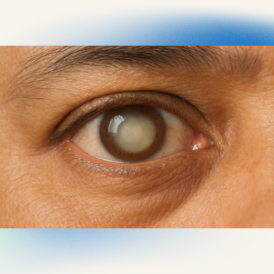
Limitations: OCT relies on light passing through the eye. If you have very thick cataracts or significant bleeding inside the eye (vitreous hemorrhage), the light may be blocked, making the scan less effective. In these cases, your doctor might recommend a B-scan ultrasound instead.
Who Needs an OCT Scan?
You should consider an OCT scan if you:
Are over the age of 50.
Have a family history of Glaucoma or Macular Degeneration.
Have Diabetes or High Blood Pressure.
Notice “wavy” lines in your vision or blurry central sight.
Are taking certain medications (like Hydroxychloroquine) that can affect the retina.
Conclusion: Don’t Wait Until Your Vision Changes
Your eyes are masters at compensating for damage. By the time you notice a “blur” or a “blind spot,” a disease may have been progressing for years. Ocular Coherence Tomography (OCT) is the most effective way to “see into the future” of your eye health.
Whether you are managing a condition or simply want the peace of mind that comes with a high-tech checkup, an OCT scan is an investment in a lifetime of clear vision.
Frequently Asked Questions (People Also Ask)
Is an OCT scan painful?
No. The scan is entirely non-contact. You will simply see a red line or a target light. There is no “puff of air” and nothing touches your eye.
Is the OCT scan safe for children?
Yes. Since it only uses light waves (no X-rays or radiation), it is 100% safe for children and pregnant people
How long does the scan take?
The imaging itself takes less than 60 seconds per eye. Including setup, you will be in and out of the testing room in under 10 minutes.
Can OCT detect glaucoma early?
Yes. OCT is one of the best tools for early detection because it can see the thinning of nerve fibers before your “visual field” (side vision) is actually damaged.
Is OCT better than a visual field test?
They are different. OCT looks at the structure of the eye (what it looks like), while a visual field test looks at the function (how you see). Most specialists use both together for a complete picture.